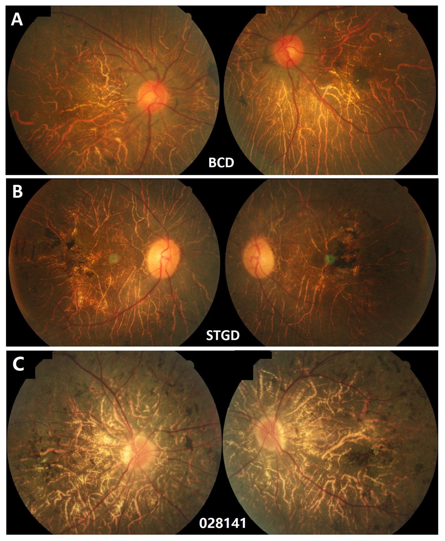
Figure 4. Fundus images of three patients with BCD or STGD.
A: Fundus images of patient 019,976 in stage 3, carrying the mutations in
CYP4V2 c.802–8_810del17insGC/p.L426F show a few crystal deposits, RPE atrophy, choroidal sclerosis, and pigment deposits.
B: Fundus images of patient 019491 with Stargardt disease (STGD) in stage 4, carrying mutations in
ABCA4 c.5196+1G>A/c.4773+1G>T [
32], display a similar fundus appearance except the crystal deposits.
C: Patient 028,141 without a mutation in
CYP4V2 in the present study shows RPE atrophy, choroidal sclerosis, and pigment deposits.
 Figure 4 of
Zhang, Mol Vis 2018; 24:700-711.
Figure 4 of
Zhang, Mol Vis 2018; 24:700-711.