Figure 3. Pedigree, fundus appearances, macular SD-OCT, ERG, and visual field images of patient 0191002. A: Pedigree of family 0191002 and segregation analysis of the homozygous mutation c.802–8_810del17insGC/ c.802–8_810del17insGC.
B: Fundus photograph of the left eye of patient 0191002 shows typical crystalline deposits in the macular region. The spectral
domain optical coherence tomography (SD-OCT) photo shows hyper-reflective material deposits in the RPE layer and RPE detachment.
C: The fundus photograph of the left eye of the mother of patient 0,191,002 shows pigment deposits and choroidal sclerosi in
the posterior pole. The SD-OCT image shows disruption of the structure of the outer retina and atrophy of the choroidal vessels.
D: The amplitude of 30 Hz flicker reaction in the electroretinography (ERG) of patient 0191002 was mildly reduced. E: Central visual field of patient 0191002 measured using a Humphrey field analyzer with a SITA 30–2 program is within normal
limits.
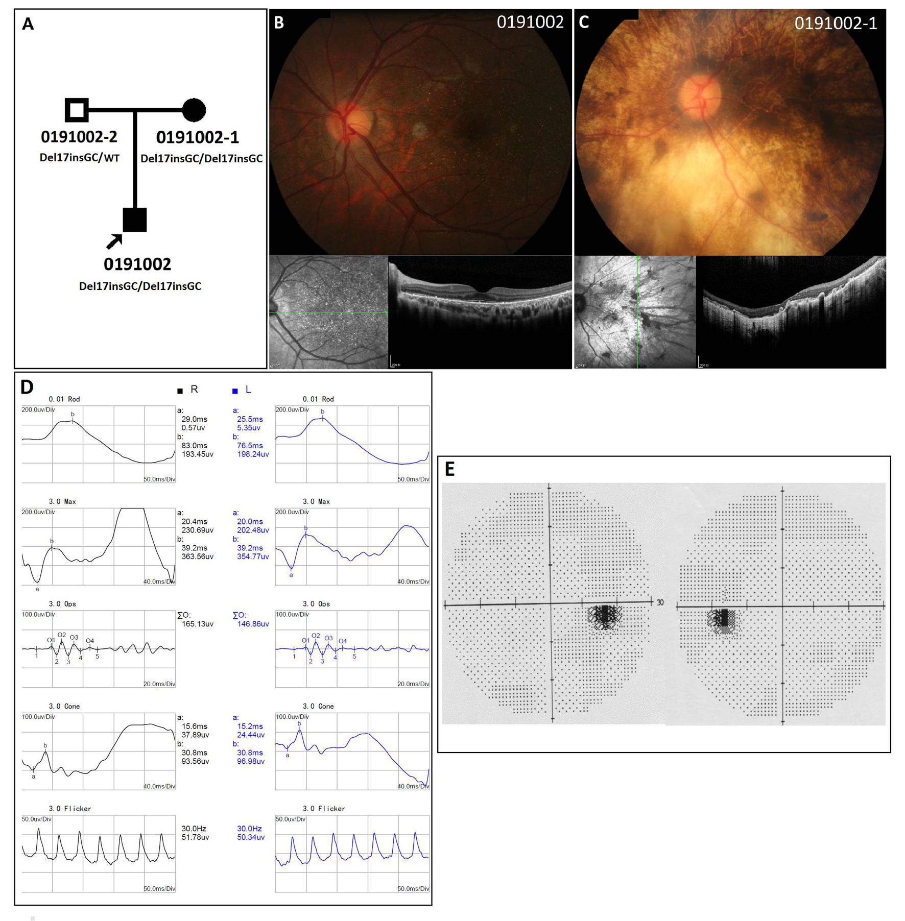
 Figure 3 of
Zhang, Mol Vis 2018; 24:700-711.
Figure 3 of
Zhang, Mol Vis 2018; 24:700-711.