Figure 2. Representative electrophoretic traces from retina and whole eye samples across different homogenization techniques. The first
peak in each trace represents a marker, the second peak denotes the 18S rRNA, and the third peak denotes the 28S rRNA.
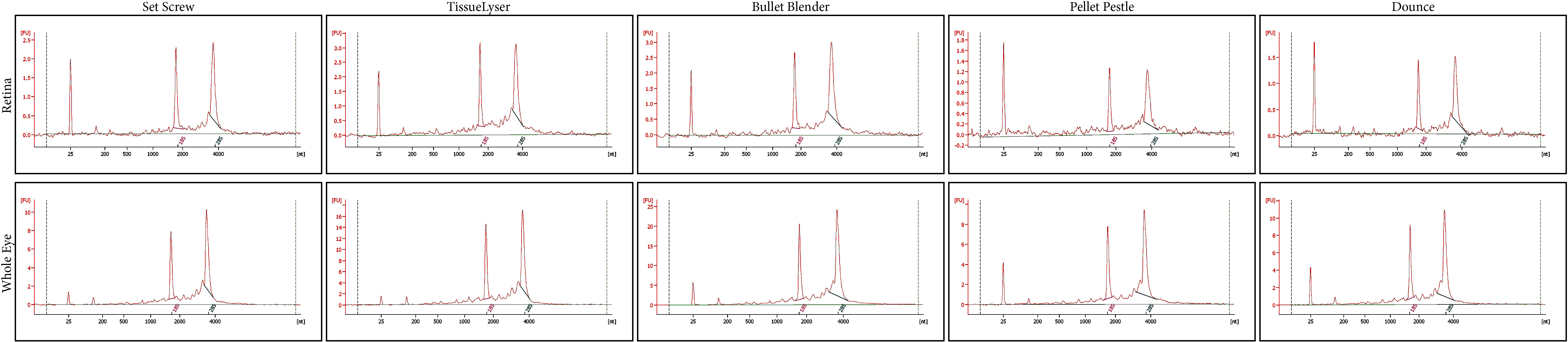
 Figure 2 of
Gooding, Mol Vis 2018; 24:690-699.
Figure 2 of
Gooding, Mol Vis 2018; 24:690-699.