Figure 2. Fundus photograph of affected individuals. A: Left and right eyes of proband (II:I) of family B presenting Drusen-like spots, attenuated vessels, pallor optic disc, and
pigmentary changes. B: Left and right eyes of proband (III:I) of family D showing diffuse pigmentary retinal degeneration, bony spicule, attenuated
vessels, and optic disc pallor.
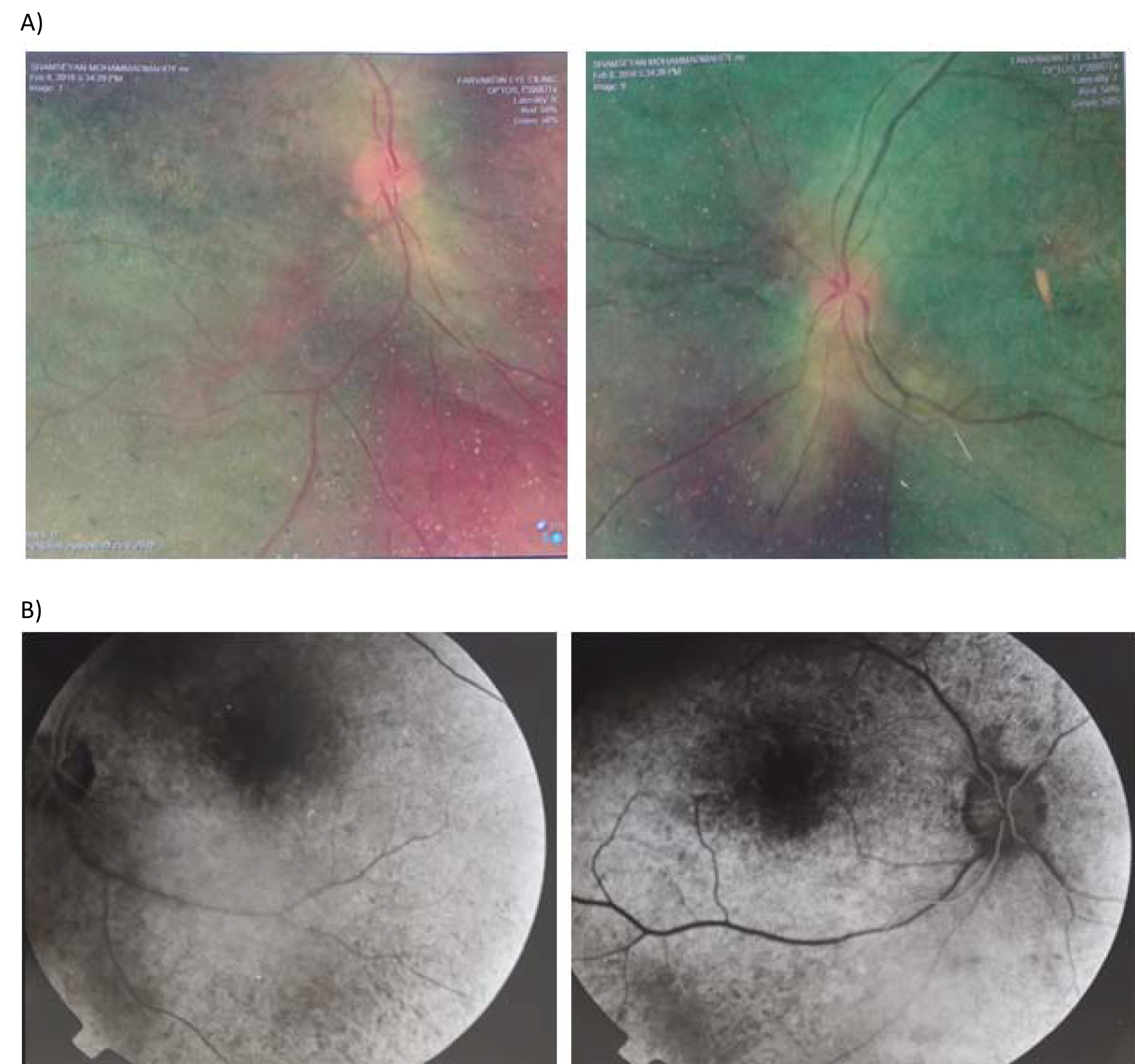
 Figure 2 of
Ravesh, Mol Vis 2018; 24:679-689.
Figure 2 of
Ravesh, Mol Vis 2018; 24:679-689.