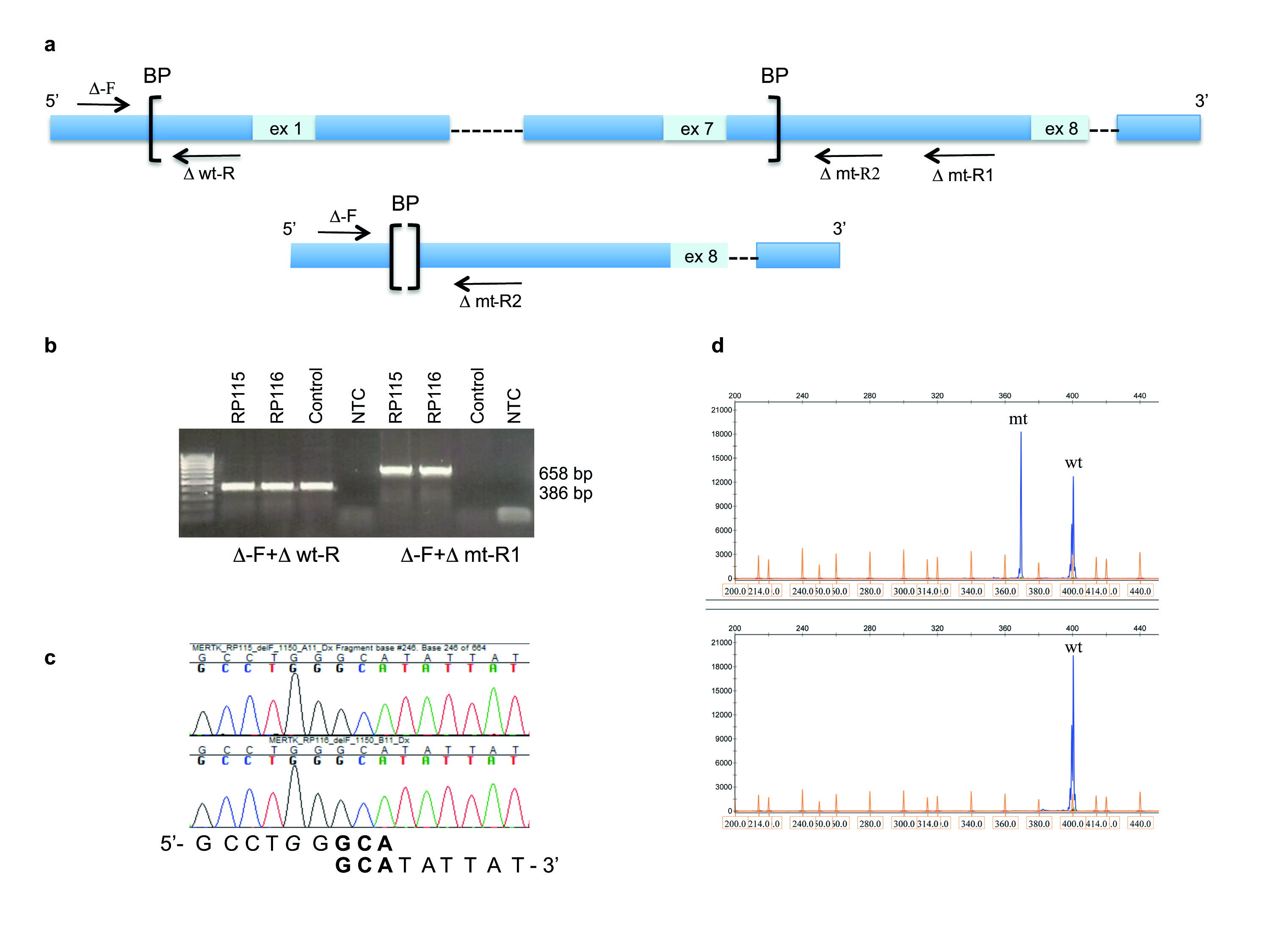
Figure 2. Characterization of genomic deletion including exons 1 to 7 of the
MERTK gene in autosomal recessive RP.
A: Localization of allele-specific primers. Primer sequences and allele-specific PCR conditions are described in Materials
and Methods and Results.
B: PCR fragments amplified with allele-specific primers were separated with agarose gel electrophoresis. Wild-type (wt) allele
(386 bp) amplified with Δ-F and Δ wt-R was detected in both affected individuals and in the unaffected control. The mutant
(mt) allele (658 bp) amplified with Δ-F and Δ-mtR1 was seen only in RP115 and RP116 and not in the control due to the presence
of a large genomic deletion.
C: Sanger sequencing shows the junction of two sequences where GCA (in bold) exists on both ends of the deletion breakpoints.
G in italics represents the retained nucleotide compared to the site of the deletion described in patients from the Faroe Islands
with retinitis pigmentosa [
11].
D: Identification of a mutant allele with fragment analysis. The upper panel shows the presence of wt and mt alleles in RP115.
The lower panel shows only the wt allele in a control sample. Fragments migrated as 370 and 400 bp despite their actual size
of 354 and 386 bp (
Table 1).
 Figure 2 of
Jonsson, Mol Vis 2018; 24:667-678.
Figure 2 of
Jonsson, Mol Vis 2018; 24:667-678.