Figure 5. Intracellular signaling involved in mediating the NaCl-induced expression of the c-Fos gene in RPE cells. The level of c-Fos
mRNA was determined with real-time reverse transcription (RT)–PCR analysis in cells cultured for 2 h in iso- (control) and
hyperosmotic (+ 100 mM NaCl) media, and is expressed as a fold of the unstimulated control. A: The following compounds were tested: the inhibitor of p38 mitogen-activated protein kinase (MAPK) activation, SB203580 (10
µM), the inhibitor of extracellular signal-regulated kinases 1 and 2 (ERK1/2) activation, PD98059 (20 µM), the c-Jun NH2-terminal kinase (JNK) inhibitor SP600125 (10 µM), and the inhibitor of PI3K-related kinases, LY294002 (5 µM). B: The following compounds were tested: the inhibitor of PLCγ, U73122 (4 µM), the inhibitor of calcium-binding proteins, ruthenium
red (Ru Red; 30 µM), the blocker of PKCα/β, Gö6976 (1 µM), the PKA blocker H-89 (1 µM), the inhibitor of Src tyrosine kinases,
PP2 (100 nM), the serum and glucocorticoid-regulated kinase (SGK) blocker GSK650394 (GSK; 1 µM), the calpain inhibitor PD150606
(100 µM), the COX inhibitor indomethacin (Indo; 10 µM), the inhibitor of mitochondrial permeability transition, cyclosporin
A (CsA; 1 µM), the reducing agent dithiothreitol (DTT; 300 µM), and the reactive oxygen species inhibitor N-acetyl-L-cysteine
(NAC; 1 mM). Each bar represents data obtained in three to 14 independent experiments using cell lines from different donors.
Significant difference versus unstimulated control: *p<0.05. Significant difference versus NaCl control: ●p<0.05.
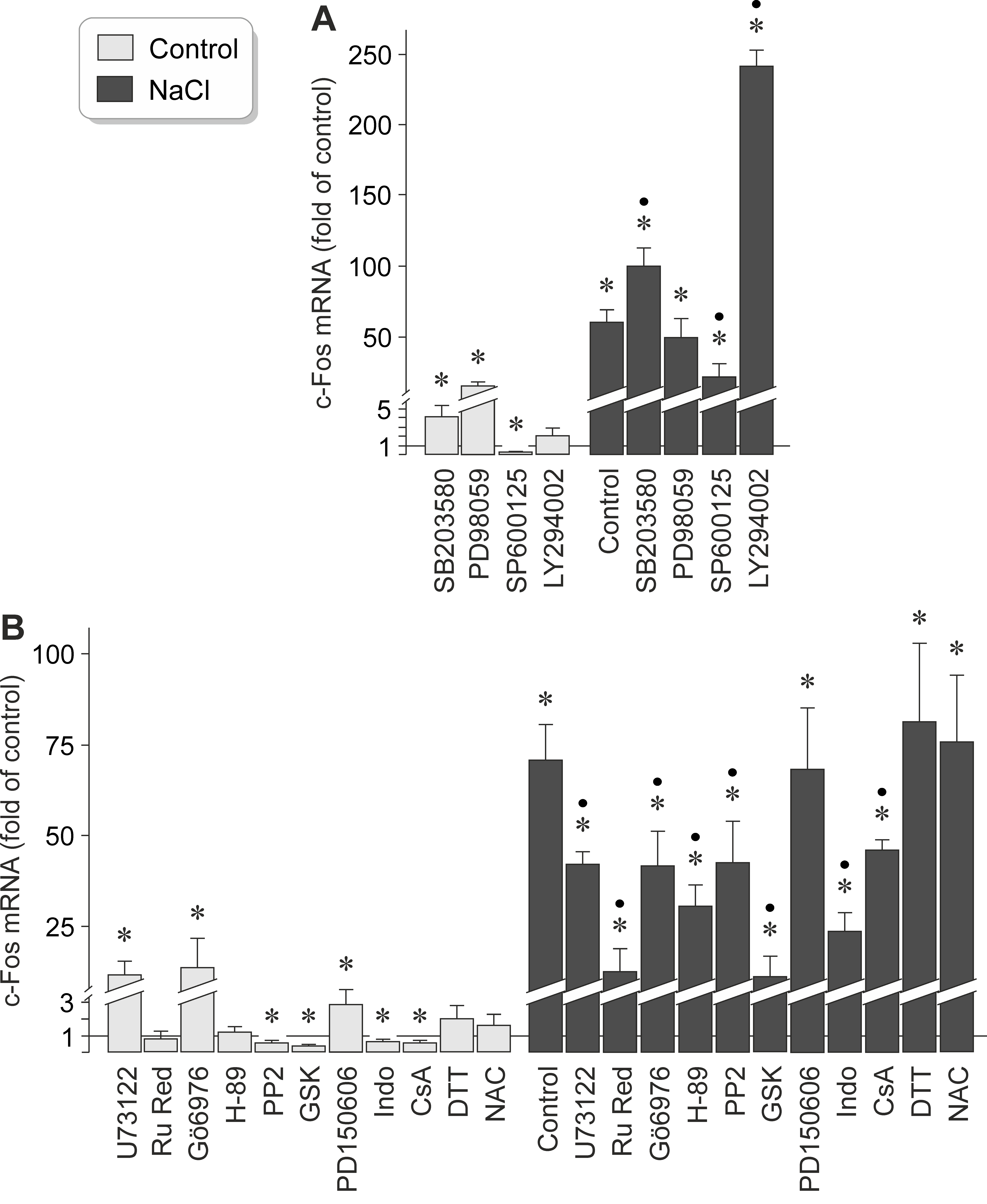
 Figure 5 of
Kleiner, Mol Vis 2018; 24:647-666.
Figure 5 of
Kleiner, Mol Vis 2018; 24:647-666.