Figure 1. Imaging of the Rhesus macaque eye pre- and post-cigarette smoke exposure. A: Hematoxylin and eosin (H&E) staining showing Rhesus macaque retina layers. B: Representative spectral domain optical coherence tomography (SD-OCT) image using the enhanced depth imaging (EDI) mode to
optimize visualization of the choroid, showing comparable retinal layers. C: SD-OCT image of the Rhesus macaque fovea region before smoke exposure. D: SD-OCT of the same eye post-exposure. Digital image segmentation of the same eye preexposure performed on the line scan
closest to the foveal center, defined as the center of the foveal pit with the greatest separation between the IS/OS junction
and the RPE layer. E: Digital images of SD-OCT images were semiautomatically segmented using the Duke Optical Coherence Tomography Retinal Analysis
Program (DOCTRAP, version 62.0), a custom image analysis software designed using MATLAB (Mathworks). F: Digital image segmentation of the same eye post-exposure. G: Fundus image of the Rhesus macaque eye. NFL, Nerve fiber layer; GCL, Ganglion cell layer; IPL, Inner plexiform layer; INL,
Inner nuclear layer; OPL, Outer plexiform layer; ONL, Outer nuclear layer; IS, Photoreceptor inner segments; OS, Photoreceptor
outer segments; RPE, Retinal pigment epithelium; CC, Choriocapillaris; C, Choroid.
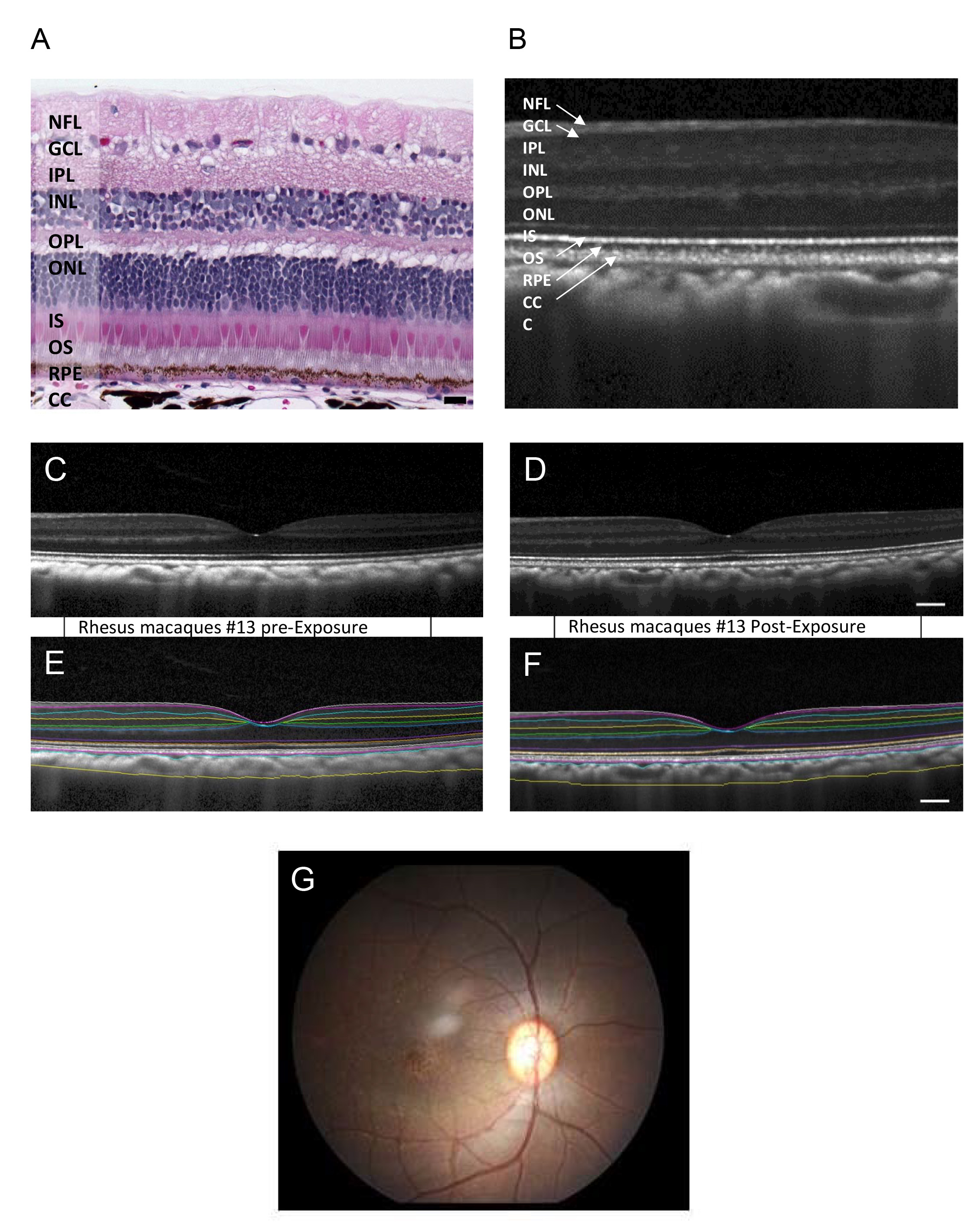
 Figure 1 of
Smit-McBride, Mol Vis 2018; 24:633-646.
Figure 1 of
Smit-McBride, Mol Vis 2018; 24:633-646.