Figure 2. Effect of bilberry extract, or Dp3G, Cy3G, and Mv3G individually on blue LED light–induced ATF4 activation in 661W cells,
a murine photoreceptor cell line, in culture. Immunoblotting shows the level of the ATF4 protein. Cells were pretreated with
bilberry extract, or delphinidin-3-glucoside (Dp3G), cyanidin-3-glucoside (Cy3G), and malvidin-3-glucoside (Mv3G) for 1 h.
After 1 h, exposure to 450 lux blue light-emitting diode (LED) light for 9 h. A: Experimental protocol in vitro. B: Typical photomicrograph and quantitative data of ATF4 protein levels using (C) bilberry extract (C), (D) delphinidin 3-glucoside, (E) cyanidin 3-glucoside, and (F) malvidin 3-glucoside. Data are the mean ± standard error of the mean (SEM; n=5 to 6). #p<0.05, ##p<0.01 versus control; **p<0.01 versus vehicle (Dunnett’s multiple comparison tests or Student t tests).
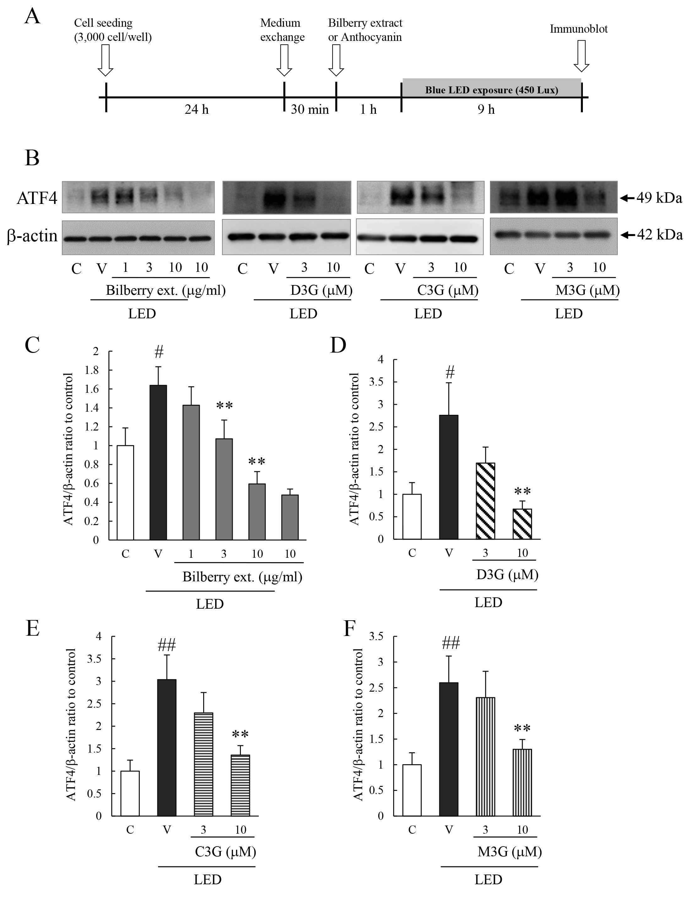
 Figure 2 of
Ooe, Mol Vis 2018; 24:621-632.
Figure 2 of
Ooe, Mol Vis 2018; 24:621-632.