Figure 3. Aberrant pre-mRNA splicing of ARL2BP as a consequence of homozygous donor splice site mutations. A: Agarose gel electrophoresis showing the reverse transcriptase (RT)–PCR products (exon 1 to exon 6) from whole blood RNA
for affected individuals from family 1 (1-II:4), family 2 (2-III:2), and a control individual (wild type, WT), as indicated.
Multiple ARL2BP transcripts were amplified (forward primer 5′-CTT TCT CCT CCG CCT CTG AT-3′, reverse primer 5′-TCA TGA GCT GAG CCT ATT GG-3′)
for individual 1-II:4, who is homozygous for the c.390+5G>A exon 5 donor site variant. RT–PCR products range in size from
429 bp to 1,035 bp due to aberrant splicing of ARL2BP. Corresponding electropherograms are shown for each transcript. A low level (7.1%) of the WT transcript was also detected.
An abnormal 419 bp transcript was amplified using the same primers in individual 2-III:2, who is homozygous for the c.207+1G>A
exon 3 donor splice site variant. The product was due to exon 3 skipping, shown in the corresponding electropherogram. B: Schematic representation of the aberrant splicing events identified in individuals homozygous for the c.390+5G>A and c.207+1G>A
variants in ARL2BP. Each splicing variant is color coded according to the arrows shown in panel A, corresponding to the transcripts detected
with RT–PCR. Exons are represented by rectangles and introns by lines. * represents the location of the variant.
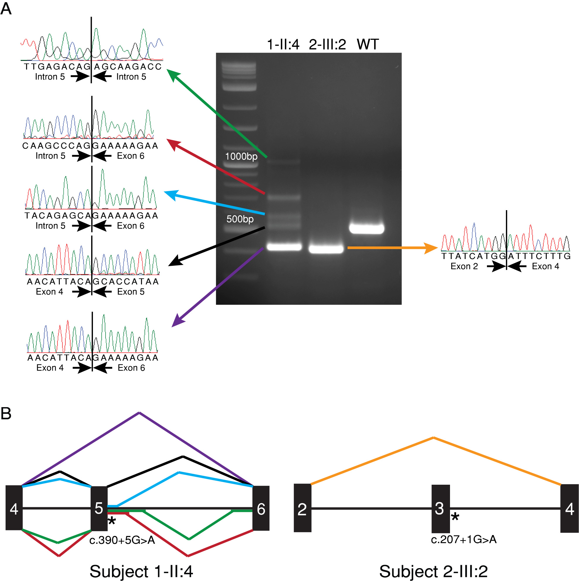
 Figure 3 of
Fiorentino, Mol Vis 2018; 24:603-612.
Figure 3 of
Fiorentino, Mol Vis 2018; 24:603-612.