Figure 2. Retinal imaging of the probands of family 1 and family 2. Family 1. A, B: Optos widefield color fundus imaging showing intraretinal bone spicule-like pigmentation mainly in the nasal retina, generalized
vascular attenuation, and optic nerve pallor. C, D: Autofluorescence imaging (Heidelberg Spectralis) showing inferiorly a reduced signal intensity in the outer macula and the
midperiphery compatible with the loss of the outer retinal structure in these areas. Conversely, the foveal region appears
intact bilaterally. E, F: Spectralis optical coherence tomography (OCT) images of the proband showing normal macular architecture. Family 2. G, H: Optos widefield color fundus imaging showing bilateral macular atrophy, and outer retinal atrophy along the vascular arcades,
with scanty bone spicule-like pigmentation mainly in the nasal retina. I, L: Optos widefield autofluorescence showing reduced signal at the posterior pole, around the arcades, and nasally, with residual
autofluorescence at the fovea in both eyes. M, N: Heidelberg Spectralis infrared and OCT images of the proband, age mid-40s, showing widespread loss of the photoreceptor
layers in both eyes, with a degree of preservation of the outer retinal structure at the fovea bilaterally. All images are
consistent with widespread loss of the outer retinal structure throughout the fundus bilaterally.
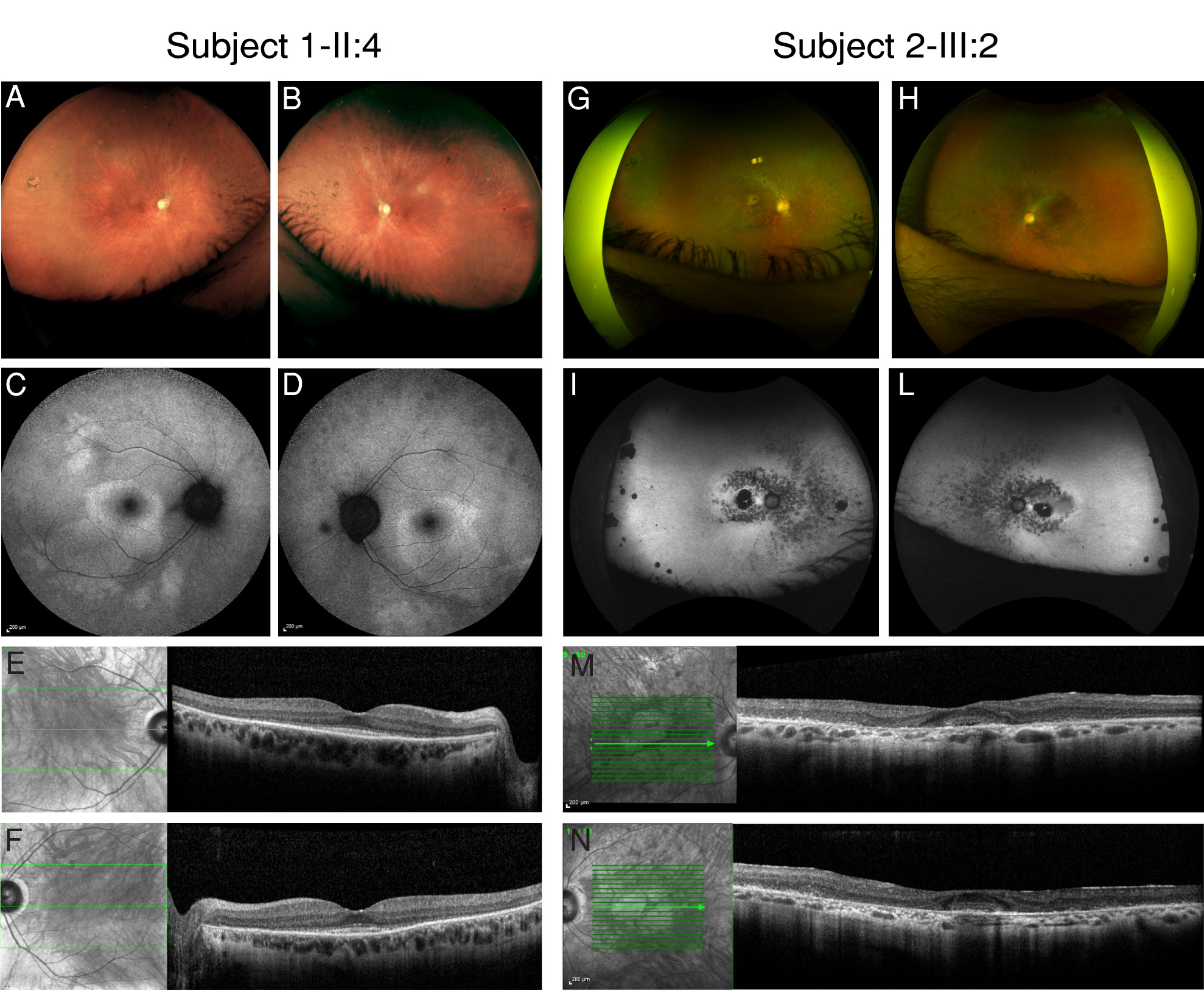
 Figure 2 of
Fiorentino, Mol Vis 2018; 24:603-612.
Figure 2 of
Fiorentino, Mol Vis 2018; 24:603-612.