Figure 3. Disruption of the N-linked glycosylation sequence in rhofl10 and rhofl7 leads to rod degeneration. A, C: Chromatograms overlaid with amino acid sequences comparing WT, rhofl10, and rhofl7 alleles, disrupting the conserved NXT consensus glycosylation sequence at N15. Red highlights deleted amino acids, while
blue highlights insertions. B, D: Confocal images of serial retinal cryosections of 5 or 6 dpf WT or heterozygous rhofl10/+ and rhofl7/+ mutants labeled with antibodies to Rho (1D1, red), rods (4C12, red), and Arr3a (Zpr-1, red) overlaid with bright-field microscopy
reveal loss of rod-specific labeling in the central retina. E: Higher magnification images of the dorsal retinal margin from WT, heterozygous rhofl10/+, or rhofl7/+ mutants showing sparse immunolabeling for Rho and rods.
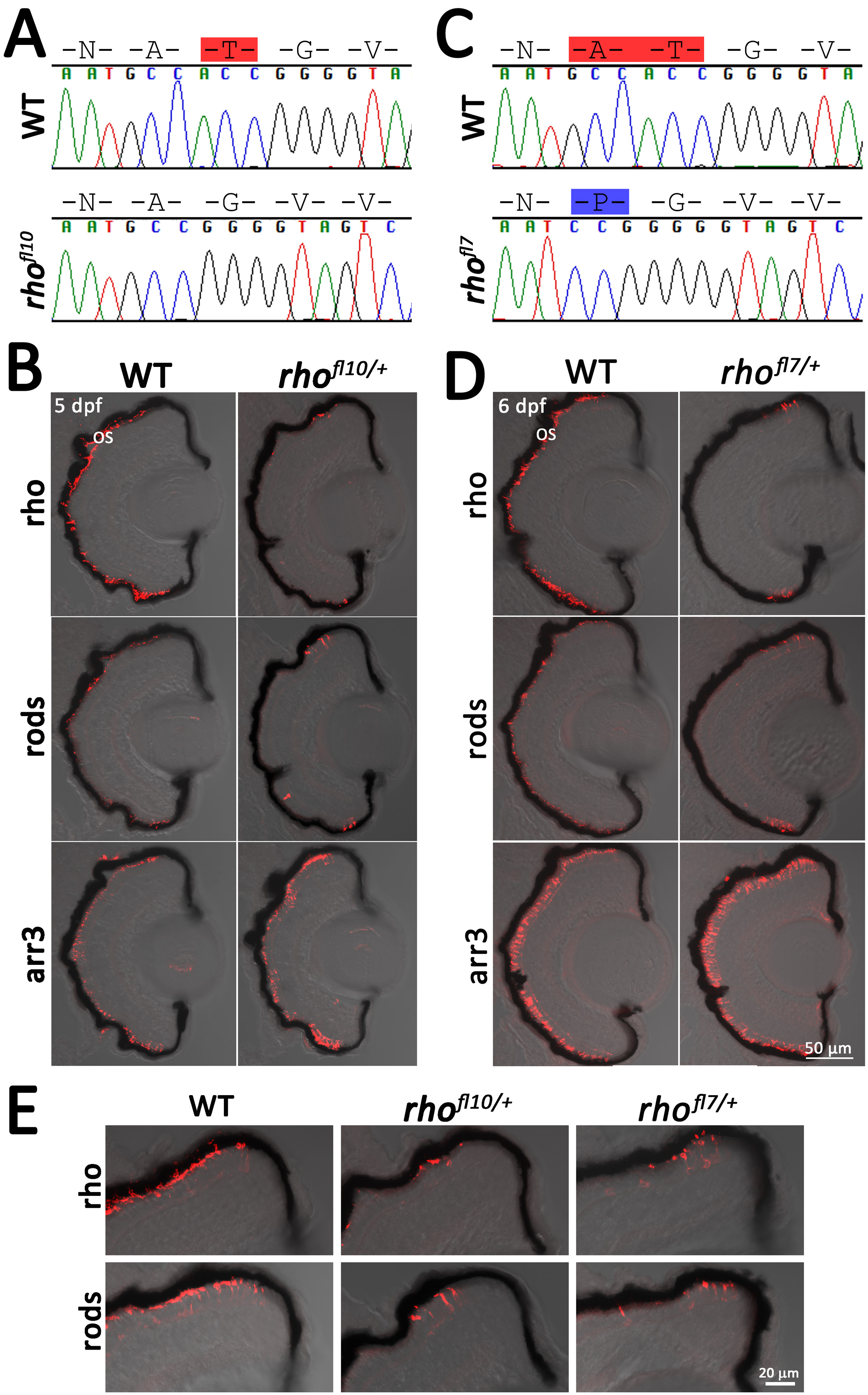
 Figure 3 of
Zelinka, Mol Vis 2018; 24:587-602.
Figure 3 of
Zelinka, Mol Vis 2018; 24:587-602.