Figure 2. Ocular manifestations of patients with mutations in COL2A1 or COL11A1. A: Photographs of the anterior segments of HM849IV2, HM849III3, HM849III4, and HM878II3. Membrane and beaded vitreous opacity
and cataracts can be seen in HM849IV2, and membrane vitreous opacity can be seen in HM849III3. Cataracts can be seen in HM849IV2
and HM849III4, while HM878II3 is normal. B: Multidirectional wide-field color photographs of HM849III3 and HM813I2, and B-scans of the left eyes of HM862II3 and HM470II1.
Retinal degeneration can be seen in HM849III3, but not in HM813I2. On the B-scans of HM862II3 and HM470II1, retinal detachment
and vitreous opacity, respectively, can be seen. C: OCT scans of HM849III3, HM862III1, and HM878II3. Posterior vitreous detachment and foveal hypoplasia (the remaining layers
in the central fovea of the macula, including the inner limiting membrane, the nerve fiber layer, the ganglion plexiform layer,
the inner plexiform layer, and the inner nuclear layer) can be detected in HM849III3 and HM862III1. The macular structure
of HM878II3 was normal.
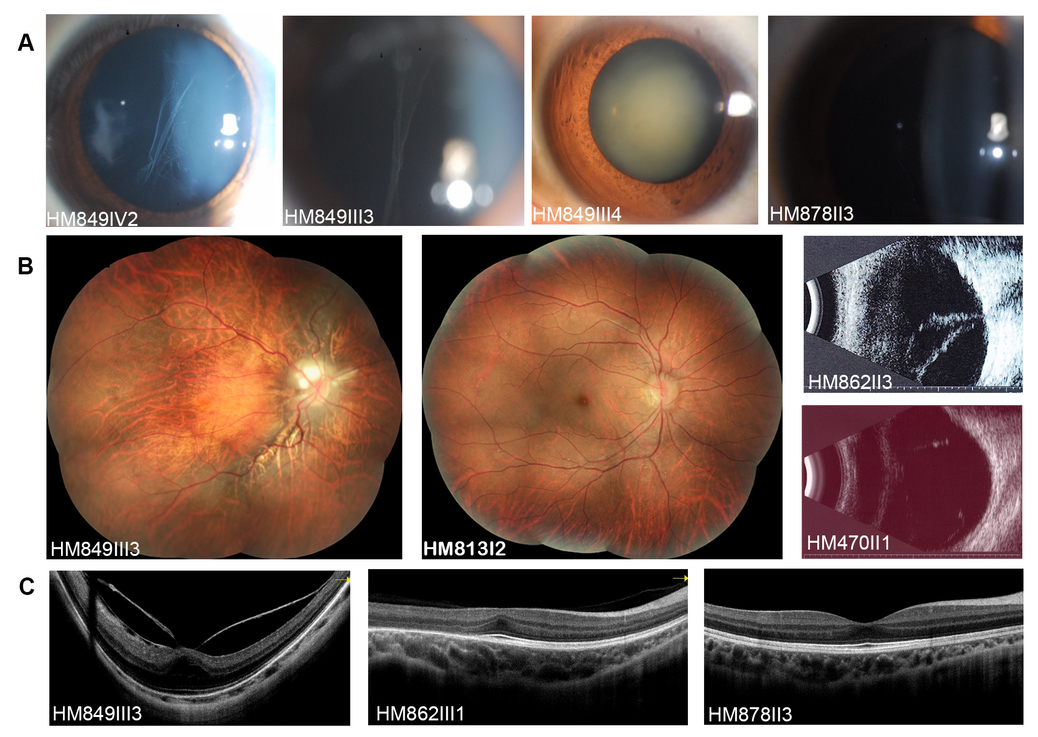
 Figure 2 of
Zhou, Mol Vis 2018; 24:560-573.
Figure 2 of
Zhou, Mol Vis 2018; 24:560-573.