Figure 1. Type I FAF phenotype: patients 14, 51, 35, and 48. Type II FAF phenotype: patients 5, 8, 11, 16, 18, 19, 21, 47, and 50. Type
III FAF phenotype: patients 4, 6, 7, 15, and 24. Negative cases at molecular testing: patients 14 and 8. Inconclusive cases
at molecular testing: patients 11, 16, 21, and 7. Patient, Pt; retinography, RG; fundus autofluorescence, FAF; infrared imaging,
IR; optical coherence tomography, OCT.
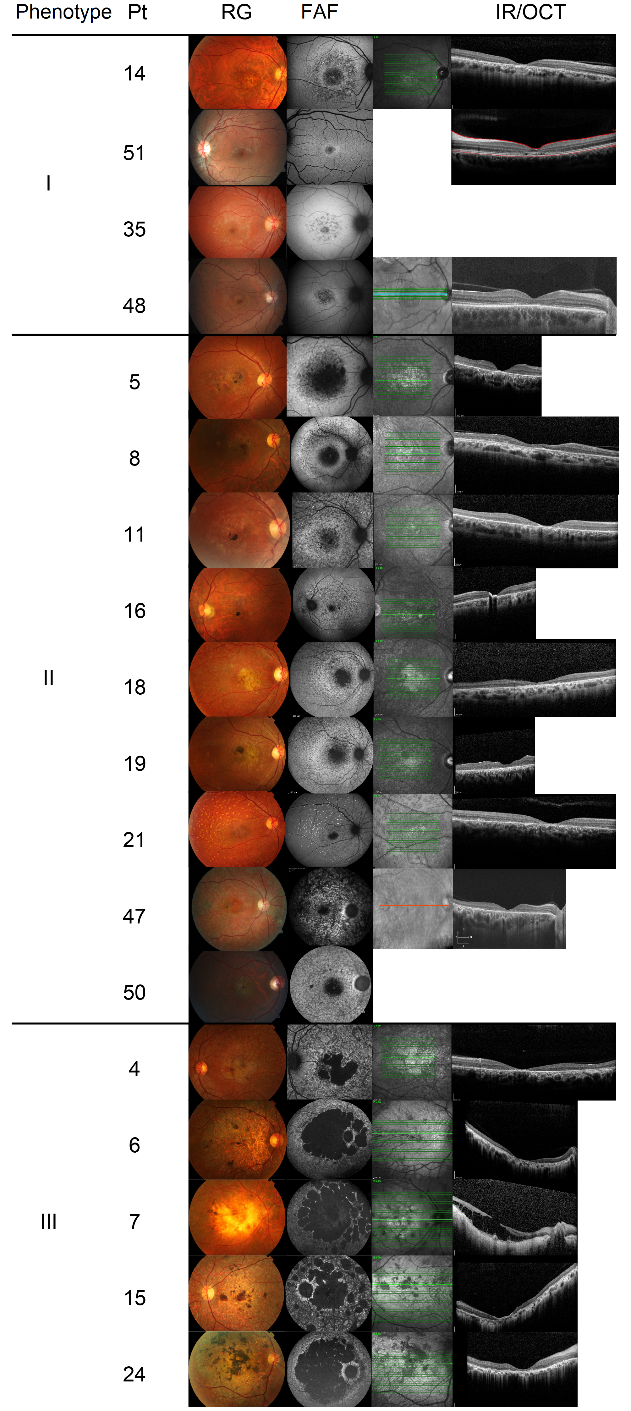
 Figure 1 of
Salles, Mol Vis 2018; 24:546-559.
Figure 1 of
Salles, Mol Vis 2018; 24:546-559.