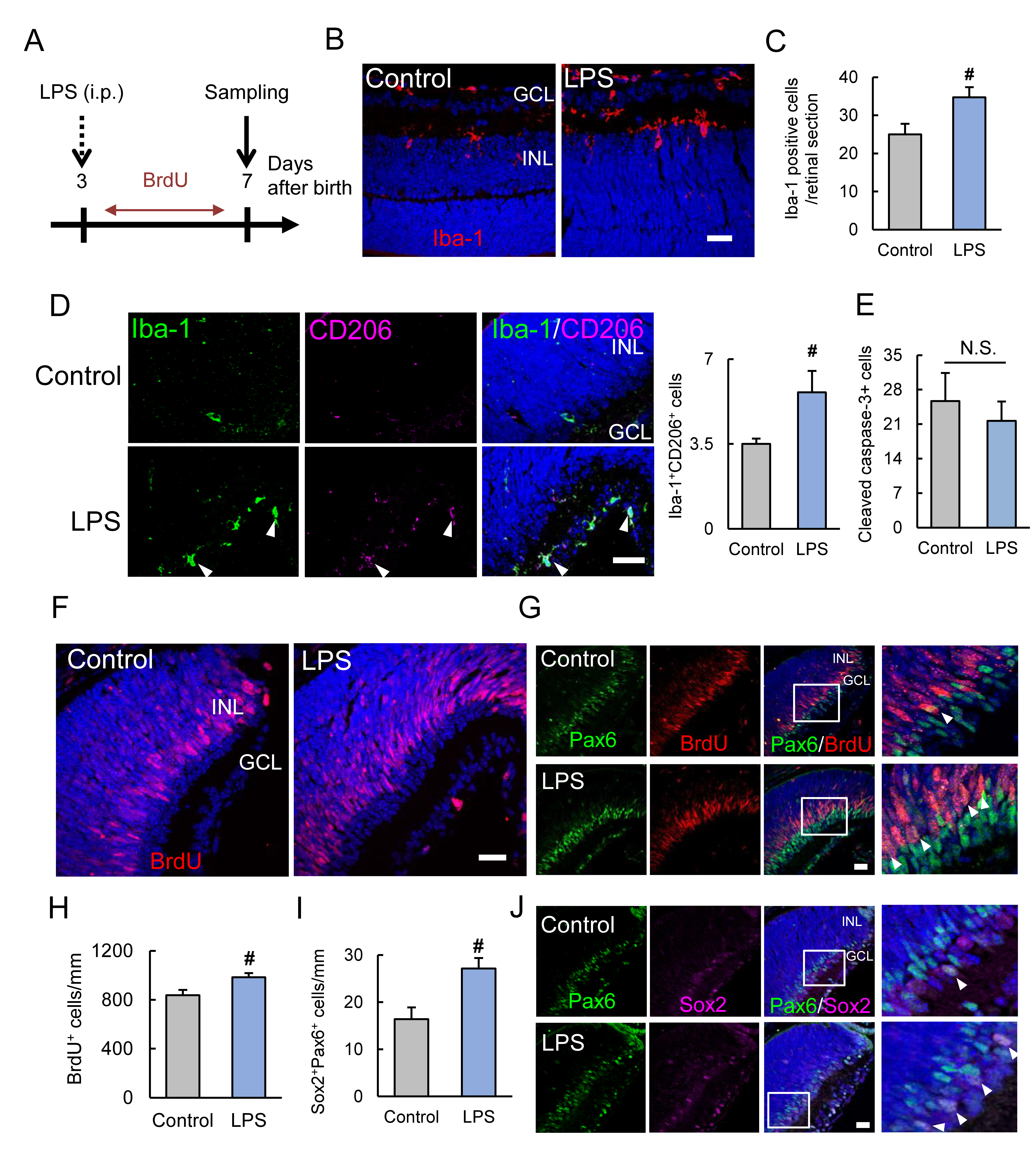
Figure 2. Systemic LPS treatment increased the number of microglia and BrdU-positive proliferative cells. A: The lipopolysaccharide (LPS) treatment scheme. Intraperitoneal injection of LPS is performed at P3, and 5-bromo-2′-deoxyuridine
(BrdU) is incorporated from P3 to P7. The retina was evaluated at P7. B, C: Immunostaining shows Iba-1-positive microglia (red). LPS treatment increases the number of microglia in the retina at P7.
D: CD206, a marker of M2 microglia, is increased by LPS treatment. E: The number of cleaved caspase-3-positive cells is not changed by LPS treatment. F–H: Immunostaining shows BrdU-positive proliferative cells (red) in the peripheral retina. LPS treatment increases the number
of BrdU-positive proliferative cells in the peripheral area. The increased BrdU-positive proliferative cells merge with the
retinal precursor cell marker, Pax6. I, J: Pax6 (green) and Sox2 (magenta) double-positive cells are increased with LPS treatment. Data are the mean ± standard error
of the mean (SEM; n=4 or 5). #; p<0.05 versus control (Student t test). Scale bar=20 μm.

 Figure 2 of
Kuse, Mol Vis 2018; 24:536-545.
  Figure 2 of
Kuse, Mol Vis 2018; 24:536-545.