Figure 6. Hyperosmotic stress increases C9 protein expression in cultured human RPE cells. A: western blot analysis of C9 protein in lysates of cultured human RPE cells and human retinas (Ret) from post-mortem donors
without reported eye disease (above). In the cases of RPE cells, 45 µg of total protein were used, while in the cases of retinas, either 40 µg (Ret 40) or 30
µg (Ret 30) of total protein were used. Middle and below: Negative controls done by omission of the first antibody (middle) and by using rabbit IgG (1:500) as the first antibody (below). B: western blot analysis of C9 protein in lysates of cells cultured in isosmotic (control) and hyperosmotic (+ 100 mM NaCl)
media for 6 h. Results of four independent experiments using cells from different donors are shown. C: Mean ± SEM C9 protein content of cytosolic extracts of cells cultured for 6 h in hyperosmotic (+ 100 mM NaCl) medium in
comparison to cells cultured in isosmotic medium (100%). The data were obtained using western blot analysis in six independent
experiments using cell lines from different donors. Significant difference versus isosmotic control: *p<0.05. D: C9 immunoreactivity in cultured RPE cells. The cells were immunolabeled with antibodies against C9 (red) and vimentin (green). Cell nuclei were stained with Hoechst 33342 (blue). Right side: Negative controls were obtained by omitting the primary antibodies. (Note that the RPE monolayer is disrupted by the fixation
procedure.) The cells were cultured for 6 h (above) and 24 h (below) in isosmotic control (left side) and hyperosmotic (+ 100 mM NaCl) media (middle). Bars, 20 µm.
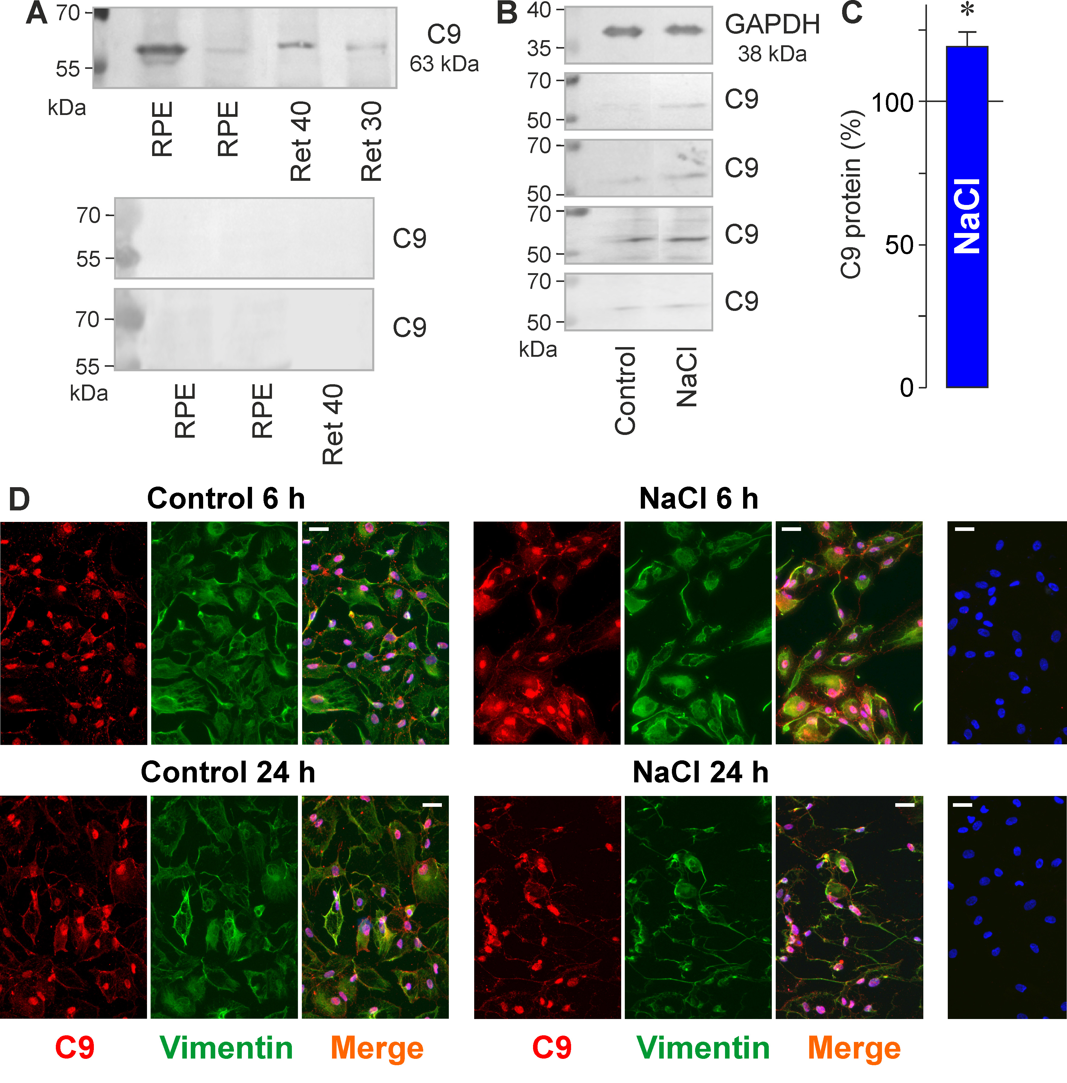
 Figure 6 of
Hollborn, Mol Vis 2018; 24:518-535.
Figure 6 of
Hollborn, Mol Vis 2018; 24:518-535.