Figure 2. Chitosan inhibits inflammation induced by IL-1β in orbital fibroblasts. The cells in medium without serum were subjected to
preincubation with chitosan (0.1 mg/ml), dexamethasone, and SP600125 for 30 min and then treated with interleukin-1 beta (IL-1β;
10 ng/ml) for 24 h. Enzyme-linked immunosorbent assay (ELISA) was used to examine the levels of IL-6 (A) and prostaglandin E-2 (PGE-2) (B) released into the medium. Another batch of cells was incubated jointly with 10 ng/ml chitosan and IL-1β (0.1 mg/ml) for
8 h. (C) Quantitative real time polymerase chain reaction (qRT-PCR) and (D) western blot analyses were conducted to evaluate the transcriptional and expression levels of COX-2. Human orbital fibroblasts
subjected to serum starvation were treated with 10 ng/ml IL-1β, and western blot analysis was used to evaluate the concentrations
of phospho-c-Jun N-terminal kinase (pJNK) concentrations after various periods of treatment. E: The JNK levels were used as the loading control. Human orbital fibroblasts subjected to serum starvation were exclusively
treated with IL-1β or were cotreated with chitosan and IL-1β for 45 min. Western blot analysis was used to detect the pJNK
quantities. F: The JNK levels were used as the loading control. Chitosan inhibits the production of IL-6 and PGE-2 induced by IL-1β. The
expression of COX-2 and PGE-2 is also downregulated by chitosan. Chitosan downregulates the phosphorylation of the JNK pathway.
Data are presented as the mean ± standard deviation (SD), **p<0.01 and *p<0.05. The experiments in this figure were repeated
three times, and similar results were obtained.
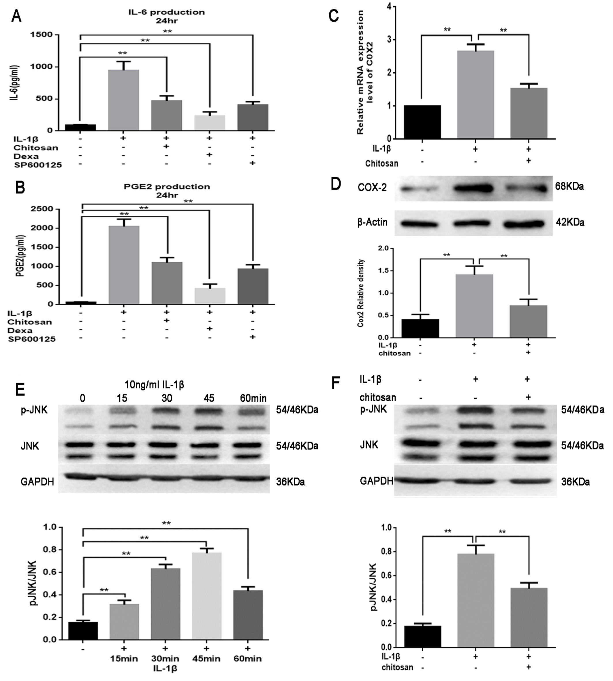
 Figure 2 of
Xiong, Mol Vis 2018; 24:509-517.
Figure 2 of
Xiong, Mol Vis 2018; 24:509-517.