Figure 1. Fundus photography of the proband. Mild macular atrophy (A) with mild peripheral bone spicule-like pigmentation in the periphery (B) at age 14 years in comparison with severe atrophic macular changes, waxy pallor of the optic disc (C), and more pronounced peripheral bone spicule-like pigmentation in the periphery (D) at age 22 years.
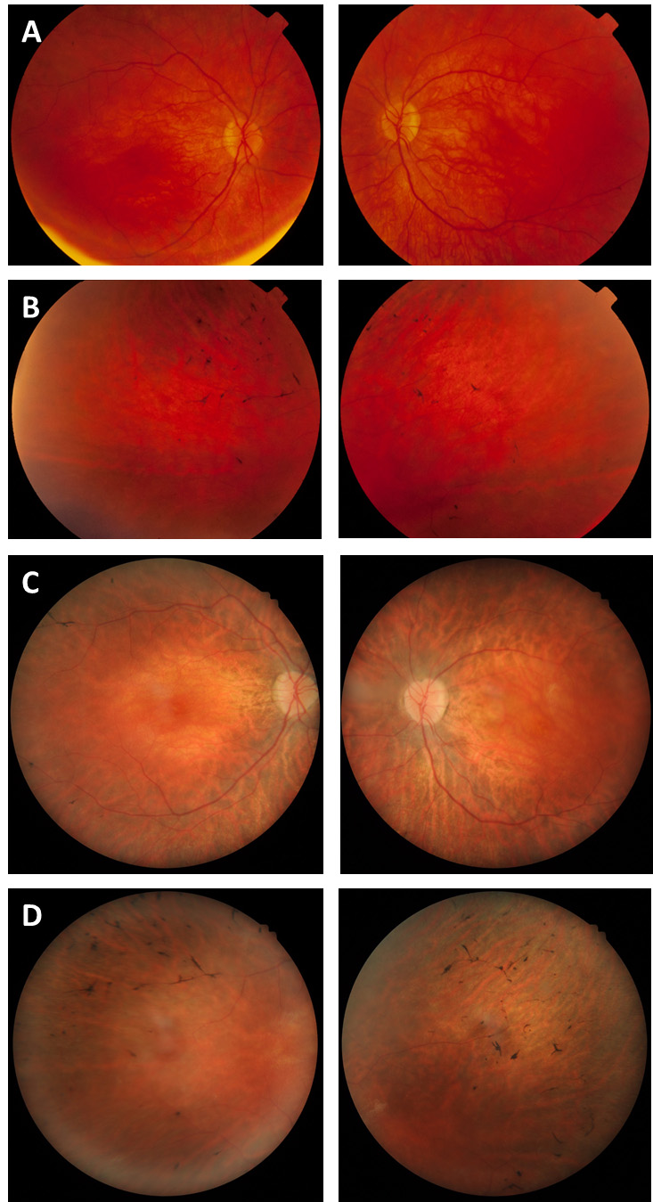
 Figure 1 of
Souzeau, Mol Vis 2018; 24:478-484.
Figure 1 of
Souzeau, Mol Vis 2018; 24:478-484.