Figure 5. Bright-field and DIC imaging of antisense and sense in situ hybridization for dscam genes in cryosections. In situ hybridization using cryosections derived from 96 hpf zebrafish retina is shown for A-D) dscama, E-H) dscamb, and C) dscaml1.A,B,E,F,I,J) In situ hybridization performed with antisense probes is shown. Arrows point to labeling in the INL and ganglion cell layer
(GCL); arrows point to labeling in the outer nuclear layer (ONL); and asterisks show labeling in the ciliary marginal zone
(CMZ). C,D,G,H,K,L) In situ hybridization performed with sense probes is shown. A,C,E,G,I,K) Photographs of antisense and sense in situ hybridization taken under bright-field conditions are shown. B,D,F,H,J,L) Images of antisense and sense in situ hybridization collected using DIC microscopy are shown. Abbreviations: DIC=differential
interference contrast; hpf=hours post fertilization; Dscam=Down syndrome cell adhesion molecule; Sdk=sidekick. Scale bar in
A=50 μm (applies to all).
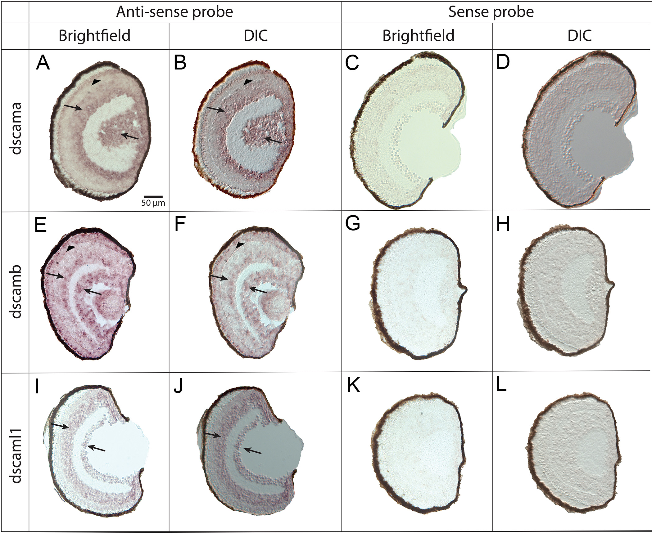
 Figure 5 of
Galicia, Mol Vis 2018; 24:443-458.
Figure 5 of
Galicia, Mol Vis 2018; 24:443-458.