Figure 5. RGR opsin in the red/green and blue cone photoreceptors of a 50-year-old donor. RGR and cone visual pigments were detected
with double-label immunofluorescent staining with DE15, and OPN1MW/LW or OPN1SW opsin antibodies, respectively. The sections
were probed first with affinity-purified DE15 and fluorescein isothiocyanate (FITC)-conjugated anti-rabbit secondary antibody.
Subsequently, the sections were incubated with the OPN1MW/LW (top panel), or OPN1SW (bottom panel) primary antibody and Alexa Fluor 568-conjugated donkey anti-goat secondary antibody. The images with 4',6-diamidino-2-phenylindole
(DAPI) counterstain showed intense labeling that revealed coexpression of RGR and cone visual pigment in the outer segments
of all red/green and blue cone photoreceptors. RGR and cone opsins were colocalized also in the inner segments of red/green
cone photoreceptors. Scale bar, 25 μm.
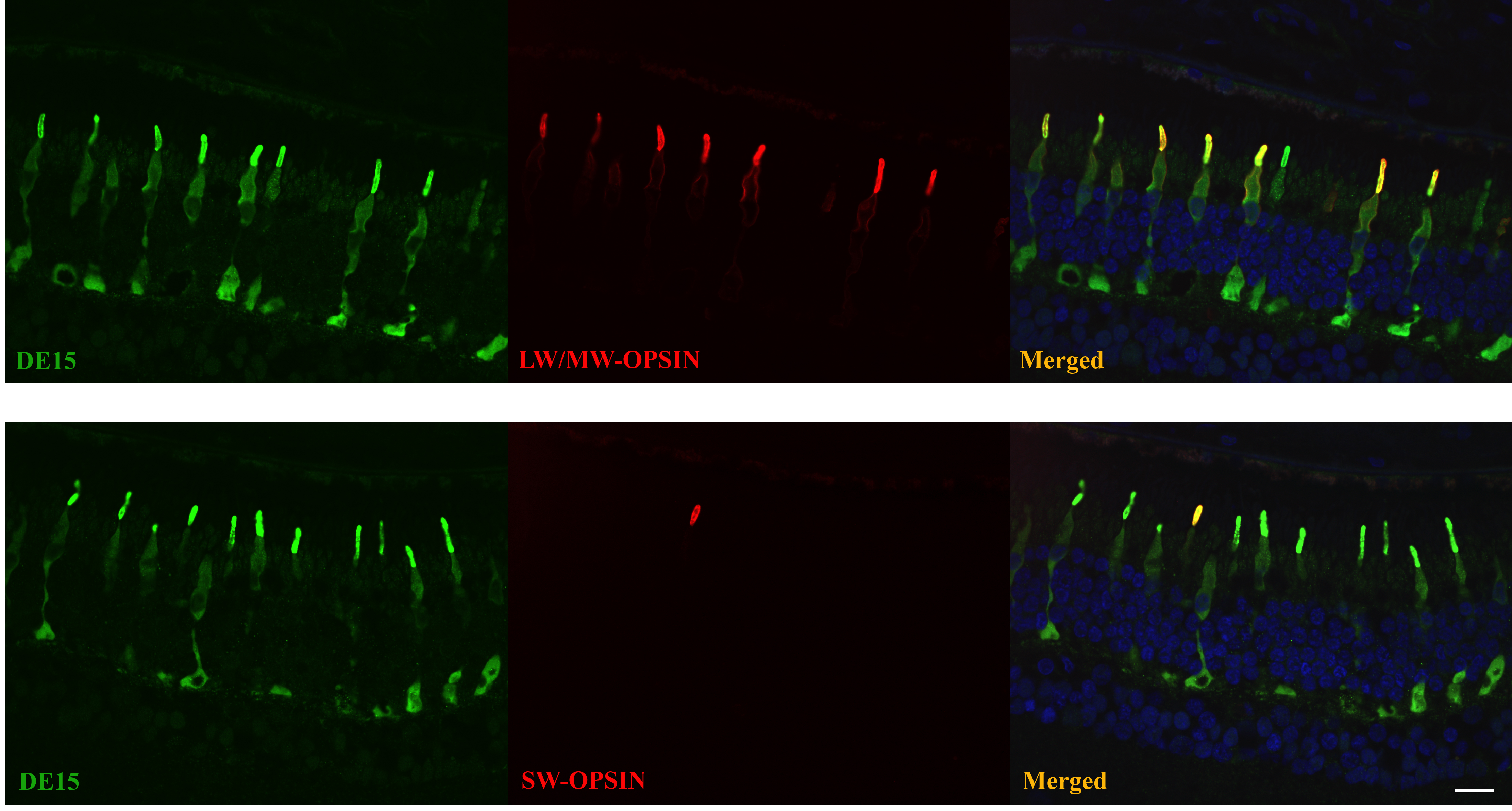
 Figure 5 of
Zhang, Mol Vis 2018; 24:434-442.
Figure 5 of
Zhang, Mol Vis 2018; 24:434-442.