Figure 3. RGR opsin in human cone photoreceptors. A: RGR opsin in cone photoreceptors of a 50-year-old human donor. Retinal sections were prepared from tissue fixed in 4% paraformaldehyde
and embedded in frozen optimum cutting temperature (OCT) compound. The sections were probed with the DE15 antibody. The tissue
section contained an intact retina still attached to the choroid. A few retinal ganglion cells (arrowheads) showed positive
immunostaining for RGR. B: The negative control slide was treated in parallel as in panel A, except that the DE15 primary antibody was omitted from the incubation buffer. C: Immunohistochemical staining of RGR-d in cone photoreceptor outer segments of an 87-year-old donor. The sections were probed
with affinity-purified DE21 antibody, which is directed against human RGR-d. The section contained the intact retina attached
to the RPE layer and choroid. RGR-d was seen in Bruch’s membrane, intercapillary regions, subcapillary regions, drusen and
basal deposits, Müller cell bodies, and cone photoreceptor outer segments (arrowheads). No counterstains were used. Scale
bars, 50 μm.
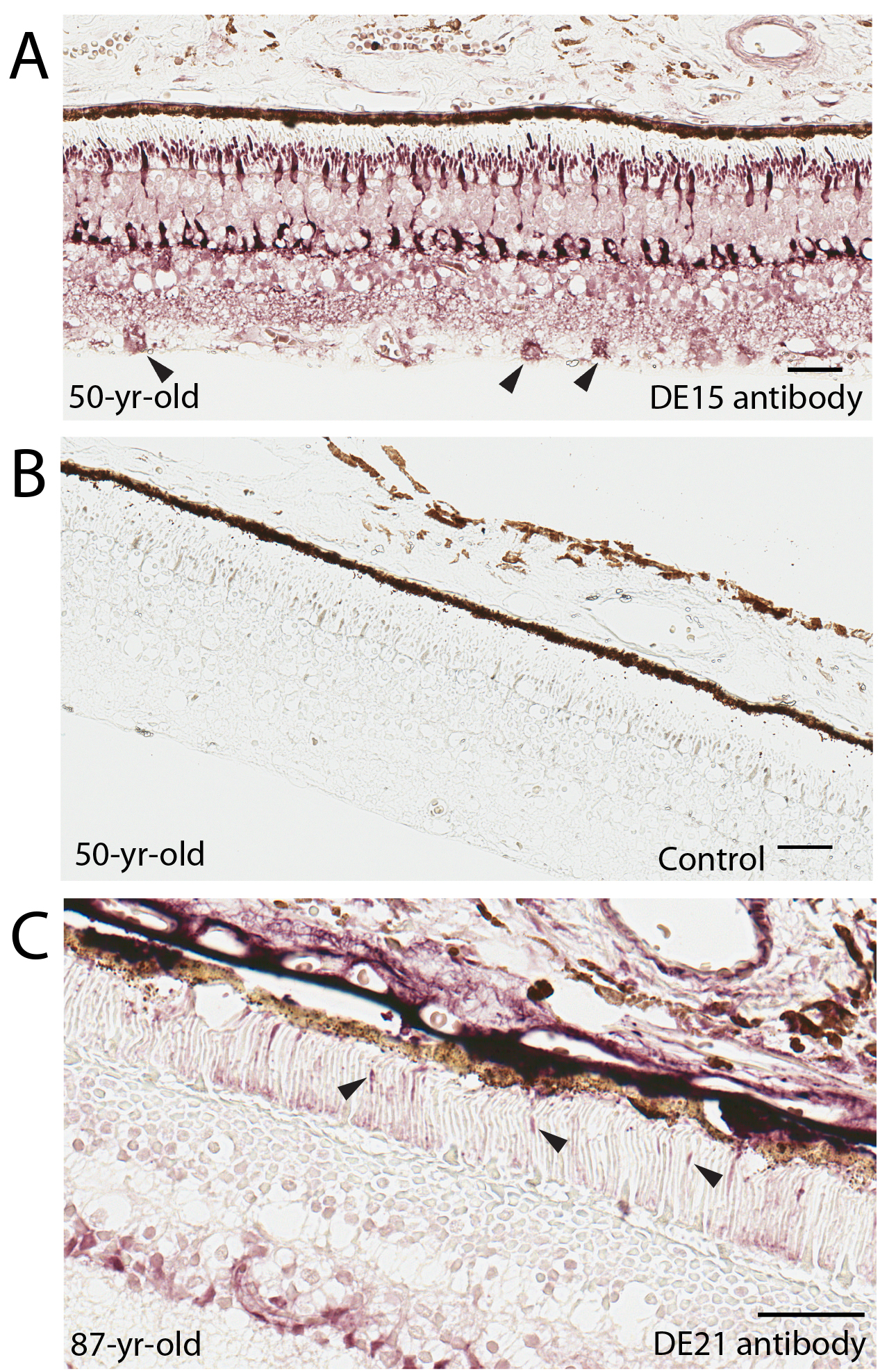
 Figure 3 of
Zhang, Mol Vis 2018; 24:434-442.
Figure 3 of
Zhang, Mol Vis 2018; 24:434-442.