Figure 2. RGR opsin in bovine cone photoreceptors. A: Retinal tissue and frozen sections were prepared from fresh bovine eyes after fixation. The tissue section was incubated
with affinity-purified DE15 antibody, and immunostaining was detected with the Vector VIP peroxidase substrate. Neuronal immunostaining
was detected throughout the cone photoreceptor and in a few retinal ganglion cells (arrowheads). Non-neuronal immunostaining
was in the RPE and in Müller cell bodies and processes. B: The negative control slide was treated in parallel, except that the primary antibody was omitted from the incubation buffer.
No counterstains were used. RPE, retinal pigment epithelium; OS, outer segment layer; ONL, outer nuclear layer; INL, inner
nuclear layer; GCL, ganglion cell layer. Scale bar, 50 μm.
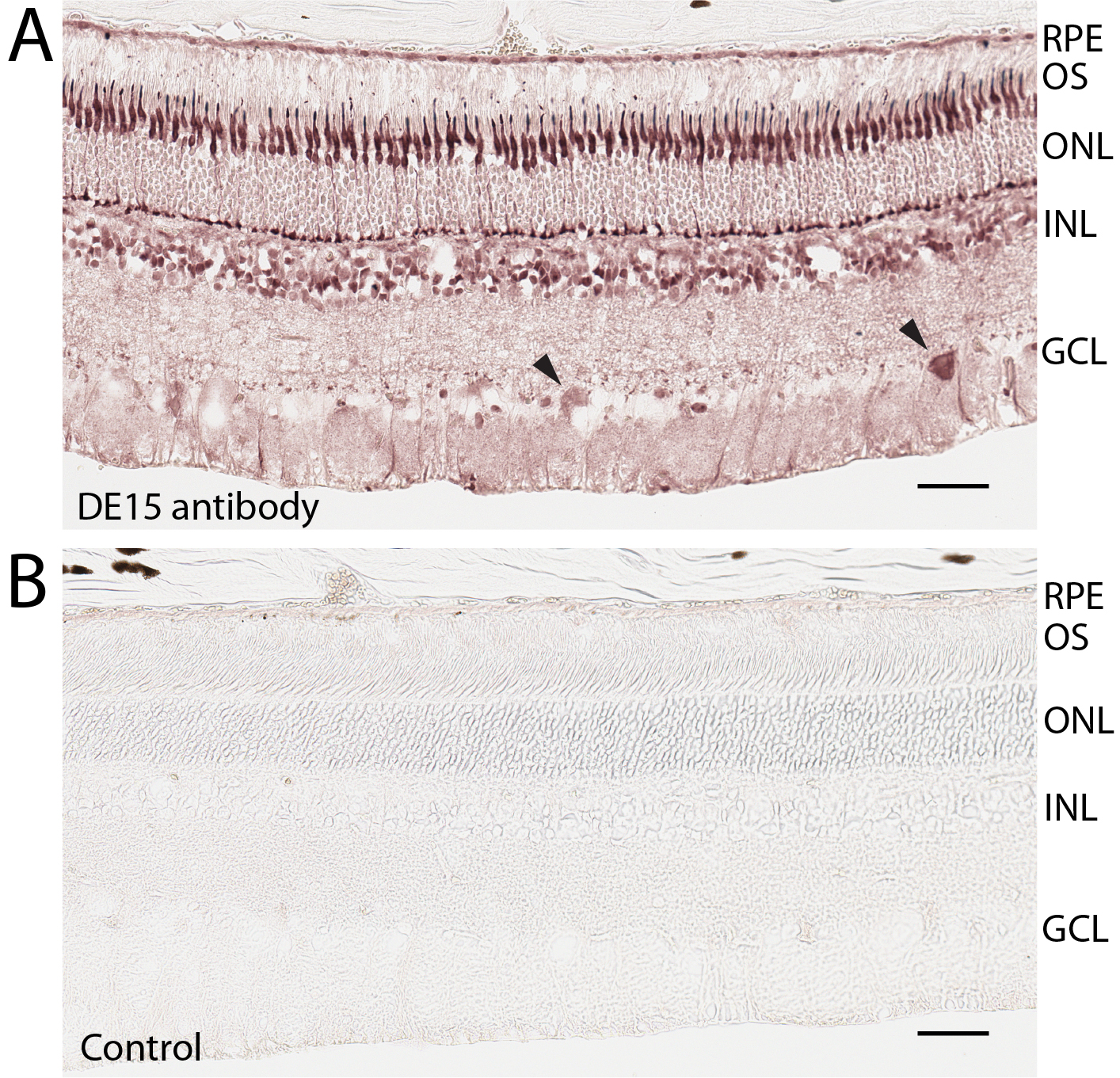
 Figure 2 of
Zhang, Mol Vis 2018; 24:434-442.
Figure 2 of
Zhang, Mol Vis 2018; 24:434-442.