Figure 3. Immunolocalization of fukutin in the 661W photoreceptor cell line. Cells were immunostained with specific antibodies against
fukutin (A, B, E; green), the Golgi marker GM130 (D, E; red), and the ER marker KDEL (G, H; red). Nuclei stained with DAPI are shown in blue (B). Enlarged views in C, F and I correspond to boxed areas in B, E and H, respectively. Fukutin immunoreactivity was found in the cytoplasm and nucleus of 661W cells, and colocalized with KDEL in
the cytoplasm (H, I, yellow), but not with GM130 (E, F). Each bar equals 20 μm, except in C, F and I: 5 μm.
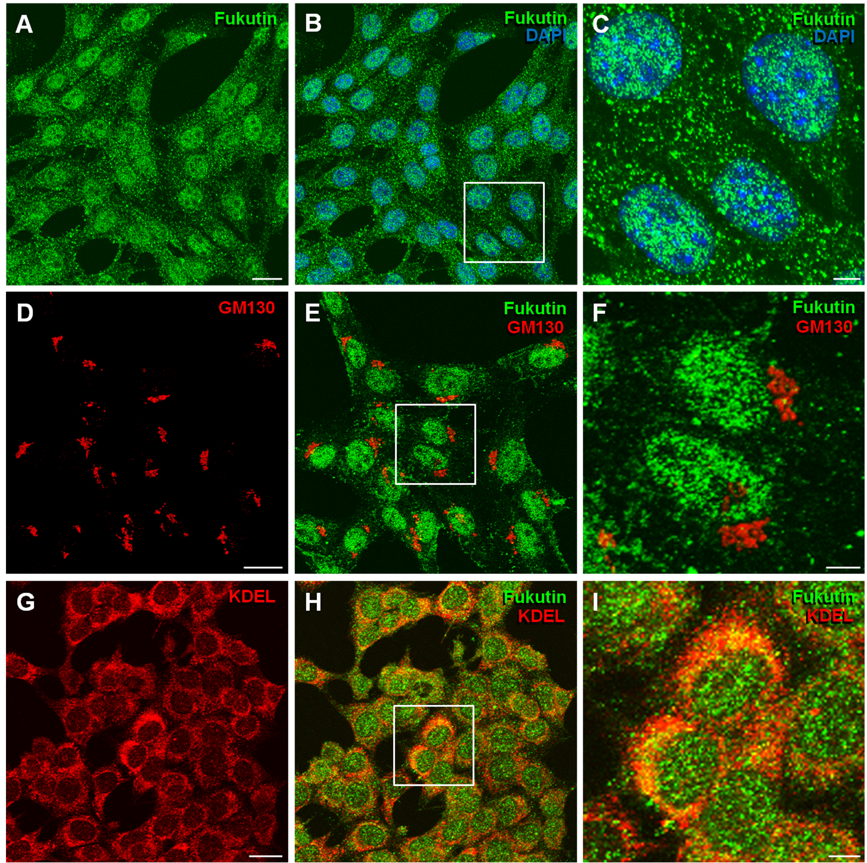
 Figure 3 of
Haro, Mol Vis 2018; 24:43-58.
Figure 3 of
Haro, Mol Vis 2018; 24:43-58.