Figure 3. Proliferative diabetic retinopathy (PDR) fibrovascular epiretinal membranes. Immunohistochemical staining for matrix metalloproteinase-14
(MMP-14) showing immunoreactivity in the vascular endothelial cells (arrows), in intravascular leukocytes, in stromal cells,
and in stromal spindle-shaped cells (arrowheads) in a membrane from a patient with active PDR (A, B, C) and in a membrane from a patient with inactive PDR (D). Notice that the membrane from the patient with inactive PDR is composed mostly of fibrous tissue. Double immunohistochemistry
for CD45 (brown) and MMP-14 (red) in a membrane from a patient with active PDR demonstrated stromal cells coexpressing CD45
and MMP-14 (arrows; E). No counterstain to visualize the cell nuclei was applied (scale bar, 10 µm).
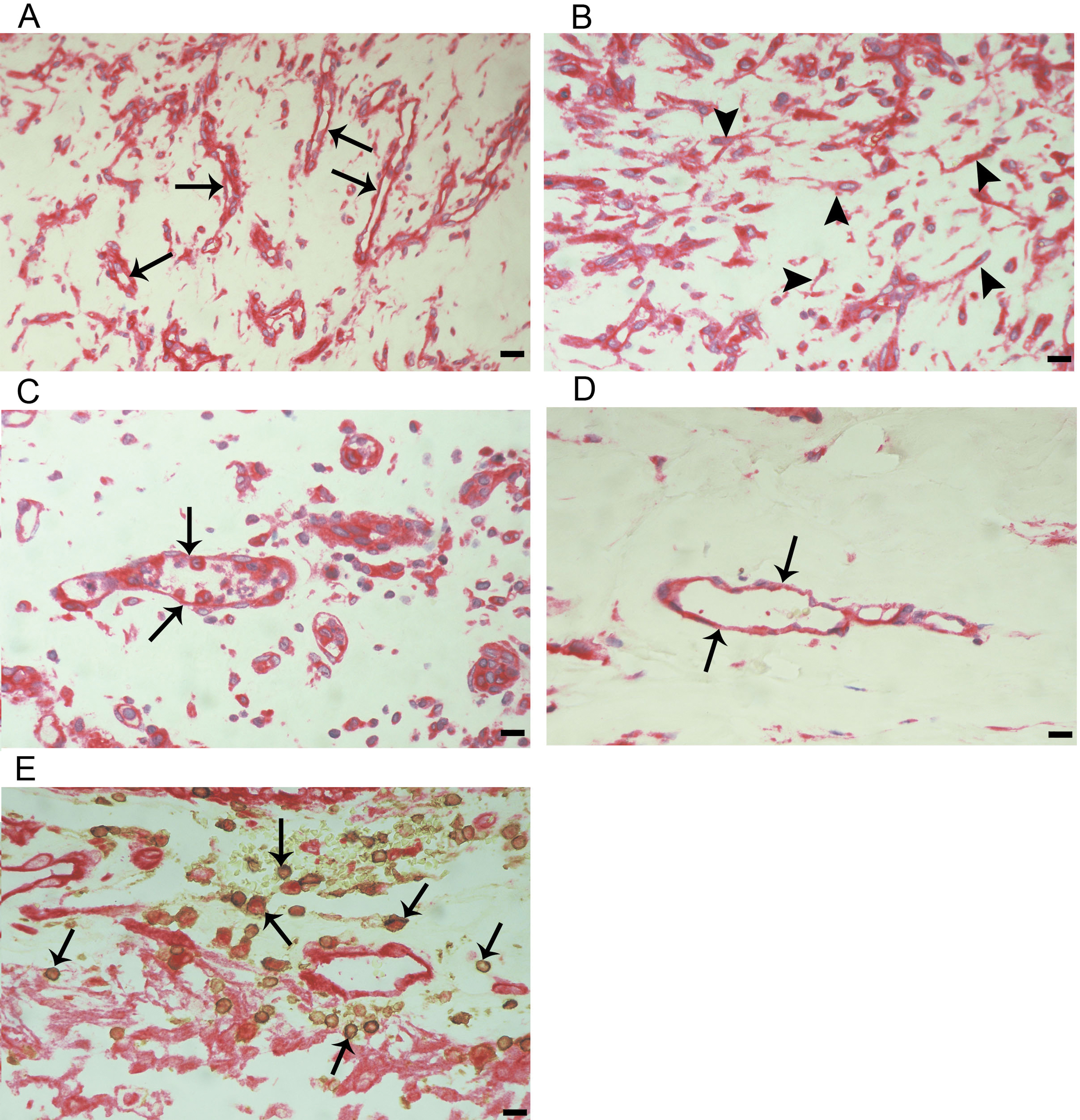
 Figure 3 of
Abu El-Asrar, Mol Vis 2018; 24:394-406.
Figure 3 of
Abu El-Asrar, Mol Vis 2018; 24:394-406.