Figure 2. Proliferative diabetic retinopathy (PDR) fibrovascular epiretinal membranes. A: Negative control slide showing no labeling. Immunohistochemical staining for CD31 showing pathologic new blood vessels expressing
this endothelial cell marker in (B) a membrane from a patient with active PDR and in (C) a membrane from a patient with inactive PDR. D: Immunohistochemical staining for CD45 showing numerous leukocytes in the stroma. E: Immunohistochemical staining for α-smooth muscle actin showing immunoreactivity in myofibroblasts (scale bar, 10 µm).
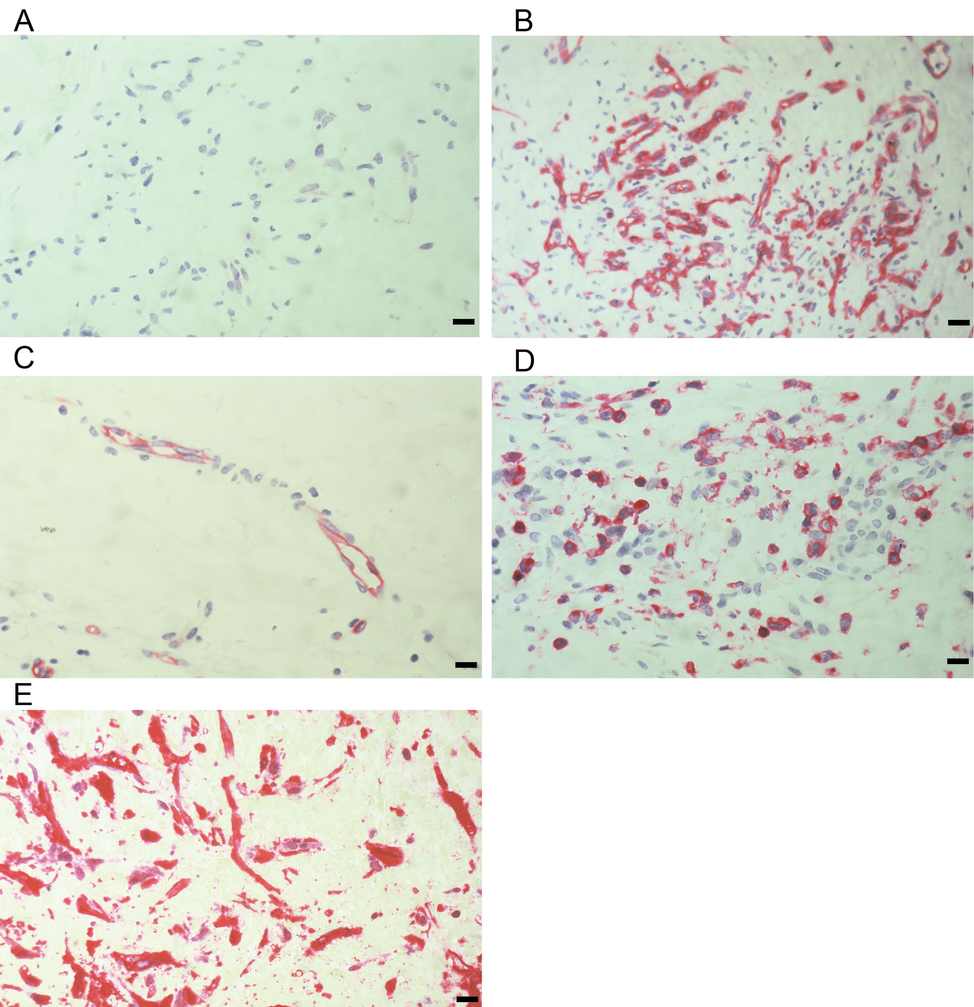
 Figure 2 of
Abu El-Asrar, Mol Vis 2018; 24:394-406.
Figure 2 of
Abu El-Asrar, Mol Vis 2018; 24:394-406.