Figure 5. Representative electron microscopic images in eyes from MCMV-infected immunosuppressed mice at day 10 post-infection or from
an uninfected control mouse. A: A negative control micrograph stained with secondary antibody alone. This shows minimal labeling in the eye of an MCMV infected
mouse. B: Immunogold staining of RIP3 in the eye of an uninfected control mouse. This shows sparse immunogold-RIP3 particles in the
choroid and RPE cells. C: Immunogold staining of RIP3 in the eye of an MCMV infected mouse. Many immunogold-RIP3 particles were observed inside the
RPE cells, some of these RIP3 stained structures appeared to be mitochondria (circles). D: Immunogold staining of RIP3 in the eye of an MCMV infected mouse. Immuogold-RIP3 particles were observed in the basement
membrane of vascular endothelial cells in the choriocapillaris. E: Double immunogold staining of MCMV EA (Larger particles) and RIP3 (smaller particles) in the eye of an MCMV infected IS
mouse. RIP3 (arrows) was observed mainly in the basement membrane of the choriocapillaris between MCMV EA (circled larger
particles in the nuclei) stained endothelial cells and pericytes.
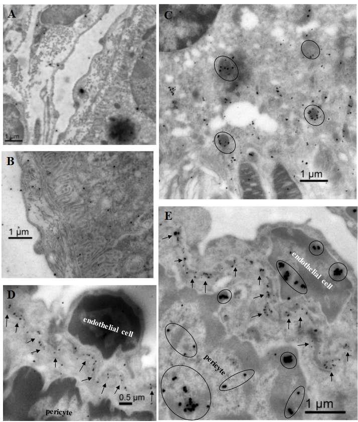
 Figure 5 of
Xu, Mol Vis 2018; 24:379-394.
Figure 5 of
Xu, Mol Vis 2018; 24:379-394.