Figure 2. Representative electron microscopic images in eye from a MCMV-infected immunosuppressed mouse at day 10 post-infection. A: Virus particles in the enlarged vesicles of RPE cells directly beneath disrupted Bruch’s membrane. B: Virus particles in enlarged vesicles of RPE cells (arrows). C: Disrupted RPE-BM (arrows).
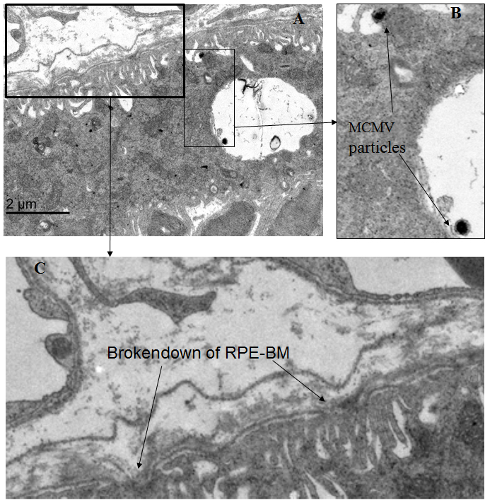
 Figure 2 of
Xu, Mol Vis 2018; 24:379-394.
Figure 2 of
Xu, Mol Vis 2018; 24:379-394.