Figure 1. Representative electron microscopic images in eyes from MCMV-injected immunosuppressed mice at day 10 post-infection. A: A negative control stained with secondary antibody alone. This shows negligible background staining in the eyes of MCMV
infected mice. B: Immunogold staining of MCMV EA showing Immunogold-labeled MCMV EA in the nuclei of some vascular endothelial cells (arrows)
and pericytes (arrow head) in the choriocapillaris. C: Adherent leukocytes in the choriocapillaris. D: Disorganization and disruption of Bruch’s membrane in areas adjacent to activated platelets which were attached to vascular
endothelia. Circles indicate disrupted CC-BM (choriocapillaris basement membrane) and RPE-BM (RPE basement membrane). E: The majority of RPE cells in the infected eye were indistinguishable from RPE cells in uninfected controls. F: An RPE cell which contained enlarged vesicles was found directly beneath disrupted Bruch’s membrane.
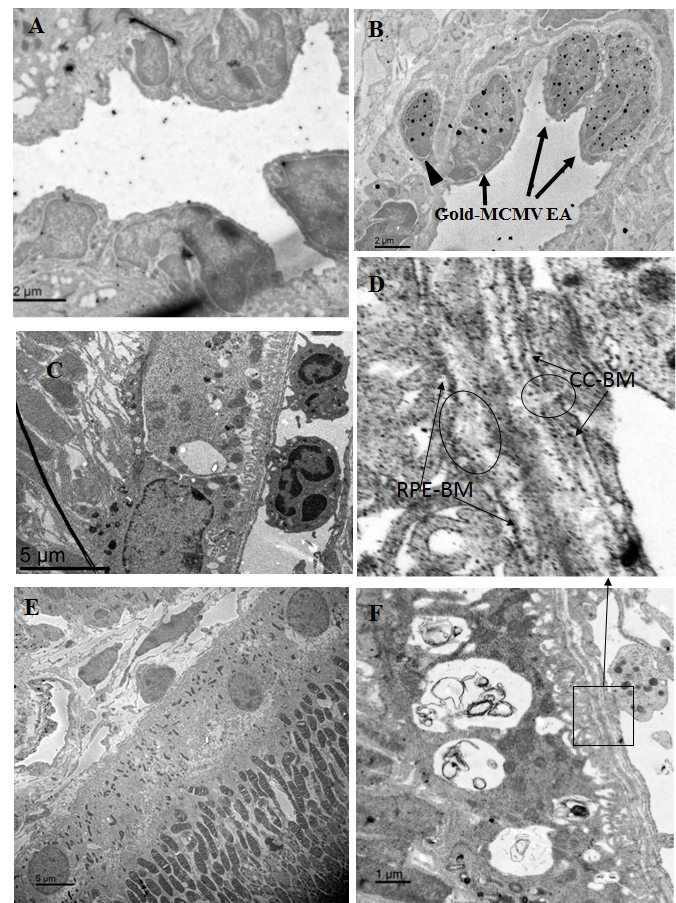
 Figure 1 of
Xu, Mol Vis 2018; 24:379-394.
Figure 1 of
Xu, Mol Vis 2018; 24:379-394.