Figure 3. Low-dose minocycline alleviated the activation of microglia in the retinal I/R injury model. The resting microglial cells
have small cell bodies and few, thin processes, while activated microglial cells are characterized by enlarged cell bodies
with numerous hypertrophied processes or amoeboid cell bodies. A–D: Whole-mounted retinas showing Iba1-positive microglia throughout the retina in the control group (A), with arrows identifying resting microglia, (B) the ischemic reperfusion injury (I/R) + normal saline (NS) vehicle group with the arrows identifying activated microglial
cells, (C) the low-dose minocycline (MC) group, and (D) the high-dose MC group. Scale bar = 100 µm. E: Quantitative estimate of the numbers of activated Iba1-positive microglia (n=7). #p<0.01,**p<0.005.
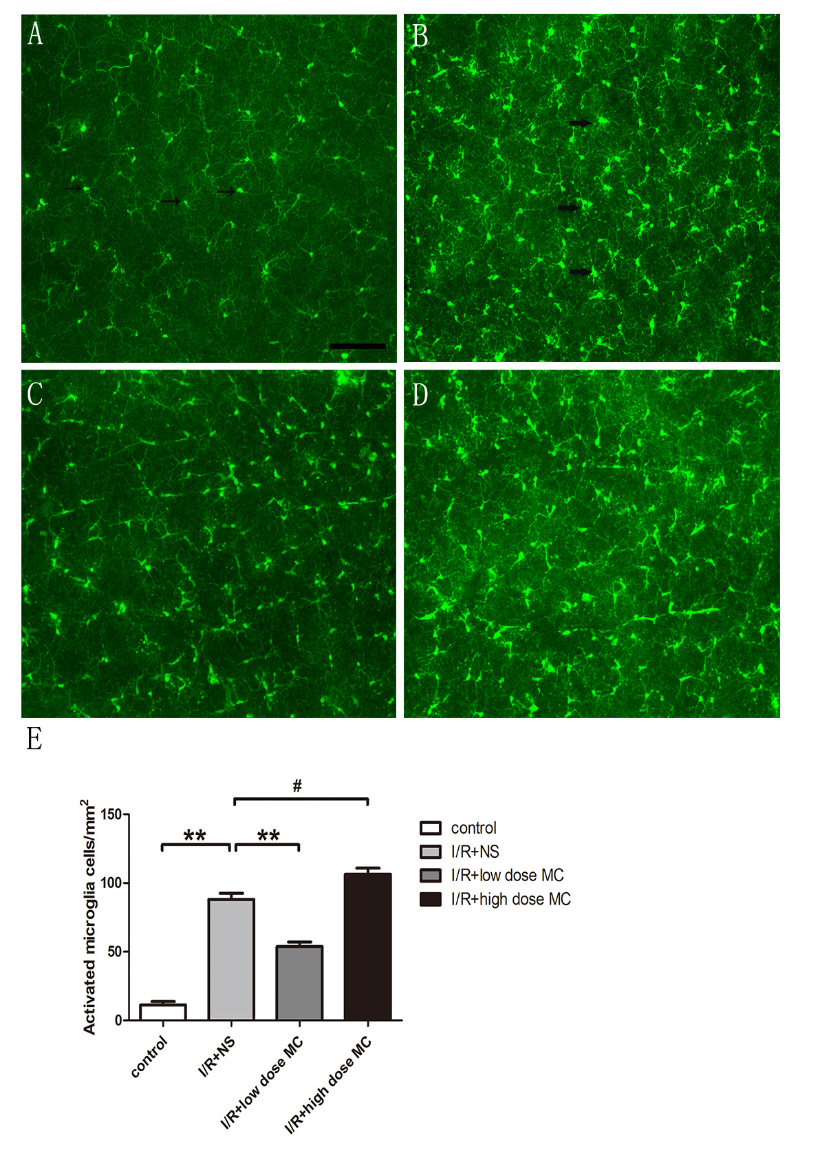
 Figure 3 of
Huang, Mol Vis 2018; 24:367-378.
Figure 3 of
Huang, Mol Vis 2018; 24:367-378.