Figure 1. Minocycline treatment reduced RGC loss in the I/R injury retina. A: Graphic showing the systemic sampling method used to count the retinal ganglion cells (RGCs). B: Whole-mounted retina immunostained with Brn3a antibody. Scale bar = 200 µm. C: The effects of test doses of minocycline (MC) on RGCs at day 4 post-ischemic reperfusion (I/R) insult, comparing each dose
of MC with the normal saline (NS) vehicle group on the number of RGCs using Brn3a immunostaining (n=5). D–G: Photomicrographs showing the mid-peripheral area of the retina labeled by Brn3a in the non-I/R injury control group (D), the I/R injury + NS vehicle group (E), the low-dose MC treatment group (F), and the high-dose MC treatment group (G). Scale bar = 50 µm. H, I: Histograms showing the percentage of surviving RGCs and the loss ratio of the RGCs (n=7). *p<0.05, #p<0.01, **p<0.005.
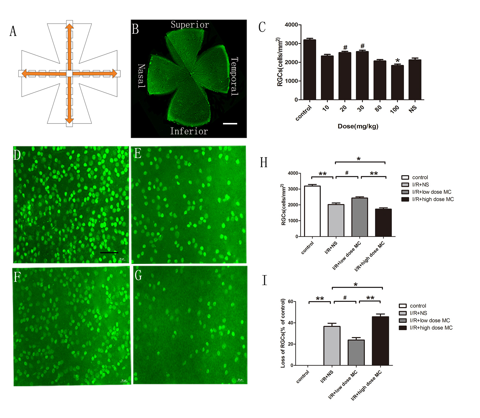
 Figure 1 of
Huang, Mol Vis 2018; 24:367-378.
Figure 1 of
Huang, Mol Vis 2018; 24:367-378.