Figure 5. HSu treatment does not statistically significantly increase the number of microglial cells in the retina. A: Retinal sections were immunostained for microglia (Iba1; red) and for reactive microglia (MHC-II; green). Nuclei were stained
with 4',6-diamidino-2-phenylindole (DAPI) ; blue). Scale bar: 50 μm. B: Translocator protein (TSPO; a marker of reactive microglia) transcript levels were assessed with quantitative PCR (qPCR),
and results are presented as the mean fold change of the control ± standard error of the mean (SEM) of 6 animals. C: The number of Iba1-positive cells was counted in the retinal sections from the control and high sucrose (HSu)-treated animals.
Results are presented as the number of Iba1-positive cells per millimeter of retina in each retinal layer and represent the
mean ± SEM. Scale bar: 50 μm.
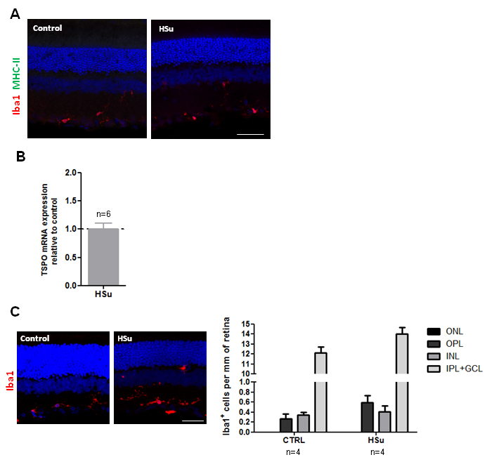
 Figure 5 of
Alves, Mol Vis 2018; 24:353-366.
Figure 5 of
Alves, Mol Vis 2018; 24:353-366.