Figure 1. Effects of exogenous PGF on morphological changes of ARPE-19 cells under hypoxia. A: The control group exhibited a cobblestone-like epithelial morphology. The hypoxia group (B) and the placental growth factor (PGF) group (C) did not show obvious morphological differences compared with the control group. D: After treatment, cells in the hypoxia+PGF group exhibited a marked transition to a more elongated spindle-like mesenchymal
morphology and were arranged in a disorderly fashion compared with the cells in the control group (A), the hypoxia group (B), and the PGF group (C). Scale bar: 200 μM.
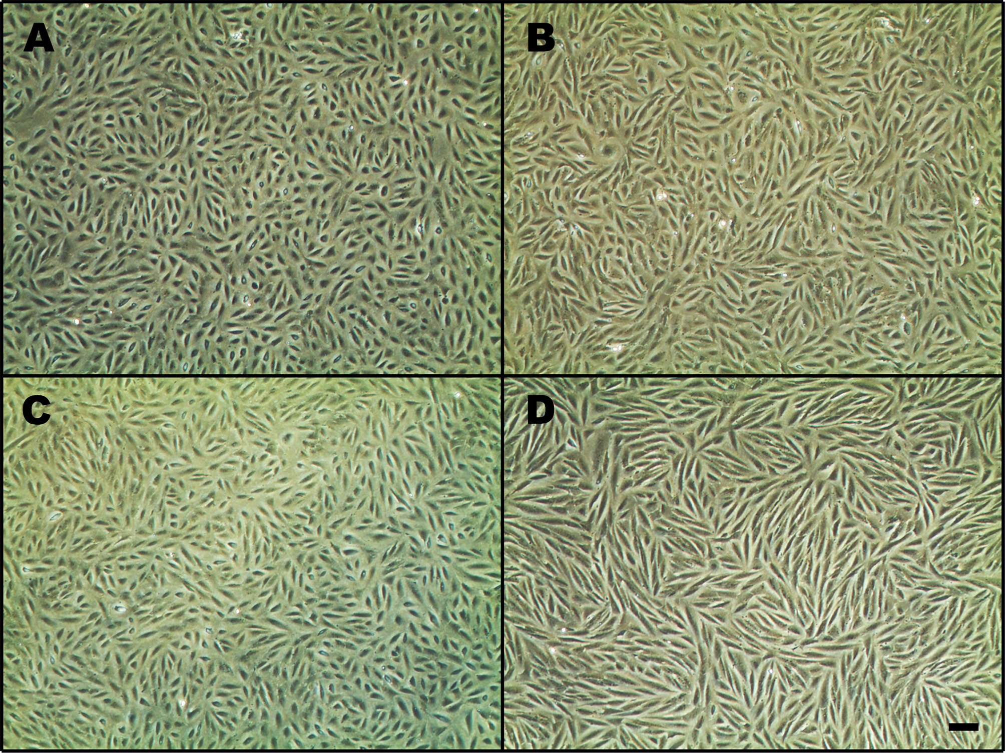
 Figure 1 of
Zhang, Mol Vis 2018; 24:340-352.
Figure 1 of
Zhang, Mol Vis 2018; 24:340-352.