Figure 5. Changes in retinal microglia. The nuclei of the microglia are labeled in green by immunofluorescence staining in the flatmounted
retina. Morphological analysis of stain-positive cells per field of view. A: Non-treated control group. B: High intraocular pressure (HIOP) model group. C: Scutellarin treatment in the HIOP model group. Images obtained at 320X magnification. The original retinal tissues that
were made into flat mounts were obtained from six eyes in each group.
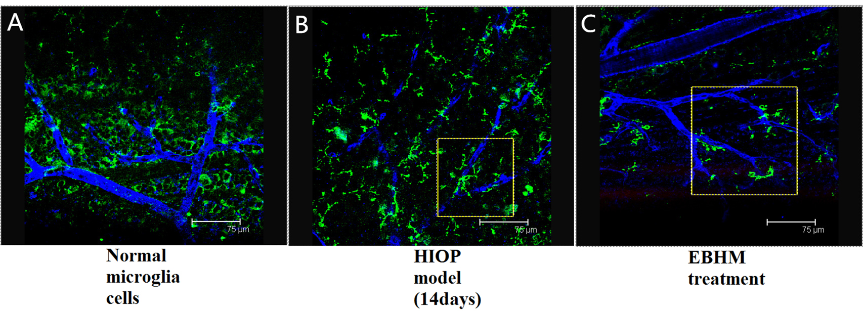
 Figure 5 of
Zhu, Mol Vis 2018; 24:315-325.
Figure 5 of
Zhu, Mol Vis 2018; 24:315-325.