Figure 2. Light micrographs of the peripheral and central ferning patterns of desert human tear film. A, B: Peripheral part of human tear film showing peripheral ferning surrounded by three layers: 1) white homogenous layer, 2)
dark brown homogenous layer, 3) granular layer with network like structure. C: “Tree branch” ferning pattern at the periphery of tear film below the peripheral layers. D, E, F: Ferning pattern of the center of the human tear film showing prominent primary, secondary, and tertiary branching. 1 = First
layer, 2 = Second layer, 3 = Third layer, F = Ferning, PF = Primary ferning, SF = Secondary ferning, TRF = Tertiary ferning.
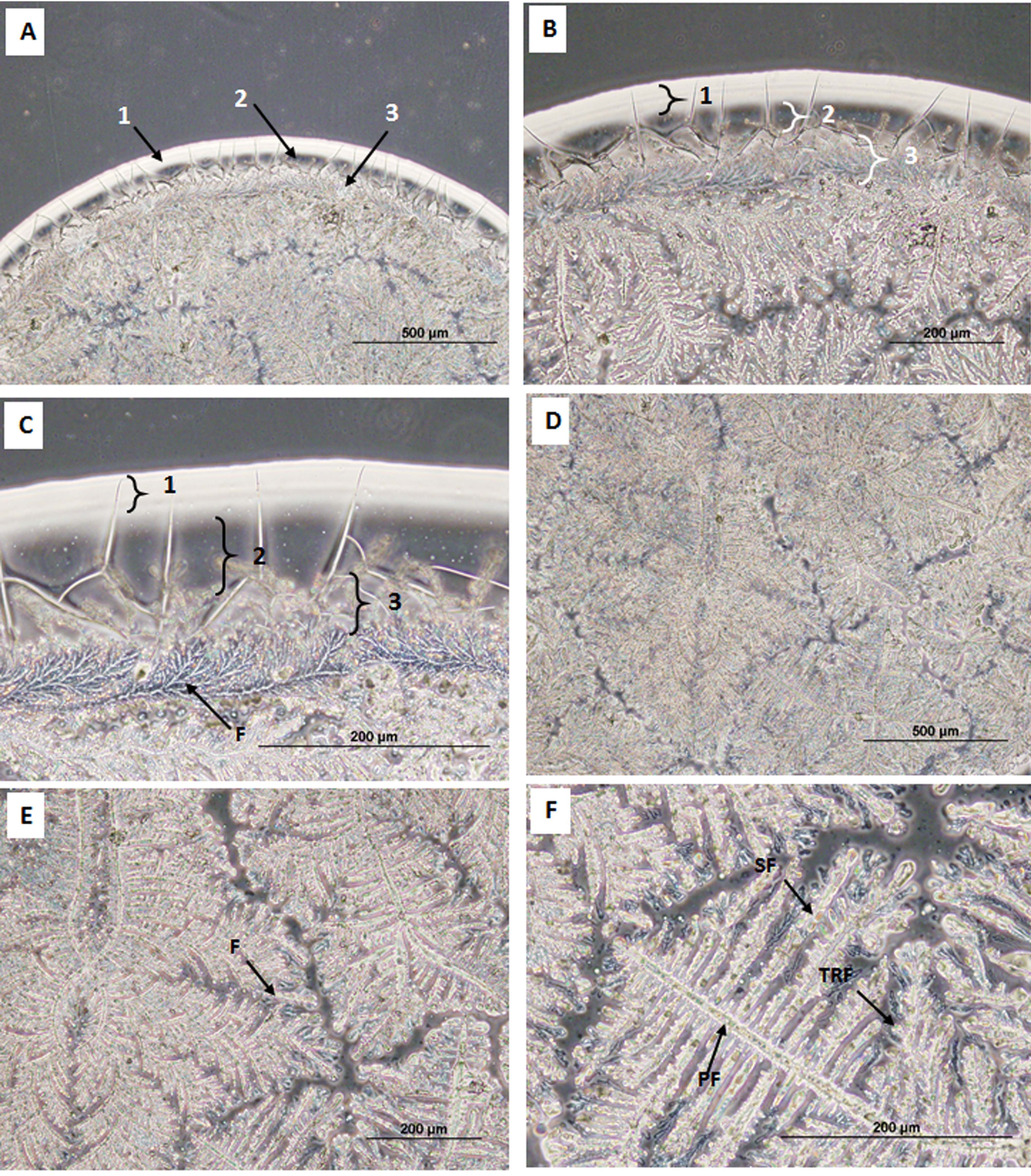
 Figure 2 of
AM, Mol Vis 2018; 24:305-314.
Figure 2 of
AM, Mol Vis 2018; 24:305-314.