Figure 2. Images of horizontal optical coherence tomography (OCT) macular scan (left column: images of the righty eye, right column:
images of the left eye). A: OCT images (patient JU#1085) show severe thinning of the outer retinal layers, with a blurred and partial disrupted ellipsoid
zone, (23 years of age). B: OCT images (patient JU#1303) reveal a continuous but blurred ellipsoid zone, (9 years of age). C: OCT images for patient JU#1303 at the age of 11 are similar to those at the age of 9. D: The outer retinal layers of the right eye are labeled in a male control without any retinal disease in his early 20s.
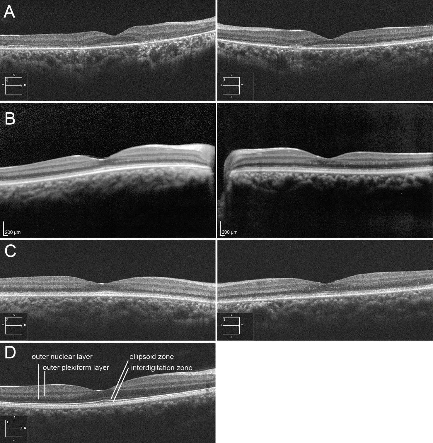
 Figure 2 of
Katagiri, Mol Vis 2018; 24:286-296.
Figure 2 of
Katagiri, Mol Vis 2018; 24:286-296.