Figure 1. Fundus images of patient JU#1085 (left column: images of the right eye, right column: images of the left eye). A: Late-phase fluorescein angiograms show hyperfluorescent rings surrounding central areas of hypofluorescence (20 years of
age). Fundus (B and C) and red-free retinal (D) images show bull’s eye maculopathy and numerous dense white dots/flecks occurring mainly outside the vascular arcades (23
years of age). E: Wide-field fundus autofluorescence images show overall low autofluorescent signals and hyper-autofluorescent rings within
the arcades (23 years of age).
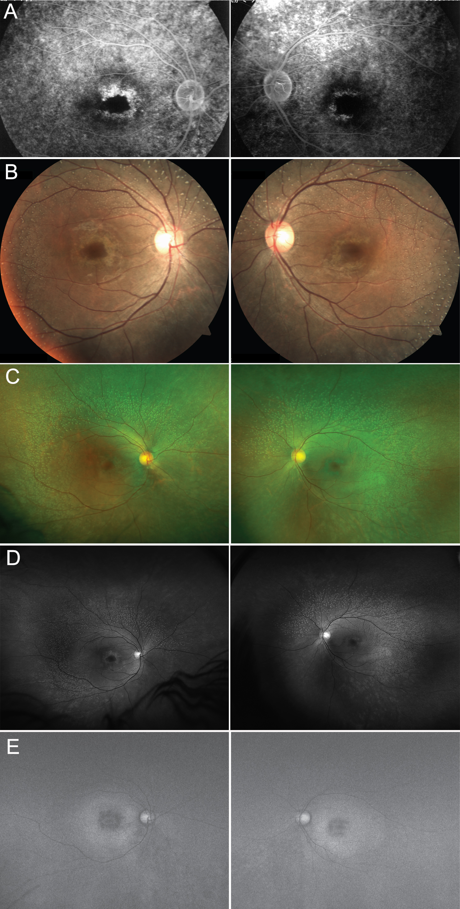
 Figure 1 of
Katagiri, Mol Vis 2018; 24:286-296.
Figure 1 of
Katagiri, Mol Vis 2018; 24:286-296.