Figure 3. Resolvin D1 restores the resolution of corneal inflammation. A: Hematoxylin and eosin (H&E) staining showed the representative histologic appearance of the healing cornea 24 h after removal
of the corneal epithelium in the control, diabetic, and resolvin D1 (RvD1)-treated diabetic mice. B: Corneas harvested 24 h after injury were homogenized and assayed for levels of myeloperoxidase (MPO) activity and tumor
necrosis factor alpha (TNF-α) and interleukin-1 beta (IL-1β) expression with enzyme-linked immunosorbent assay (ELISA; n=6).
C: Immunofluorescence staining was performed with the macrophage marker anti-F4/80 (green fluorescence) and the M2 macrophage
marker anti-CD206 (red fluorescence) 48 h after removal of the corneal epithelium. Data are given as the mean ± standard deviation
(SD); *p<0.05, ***p<0.001.
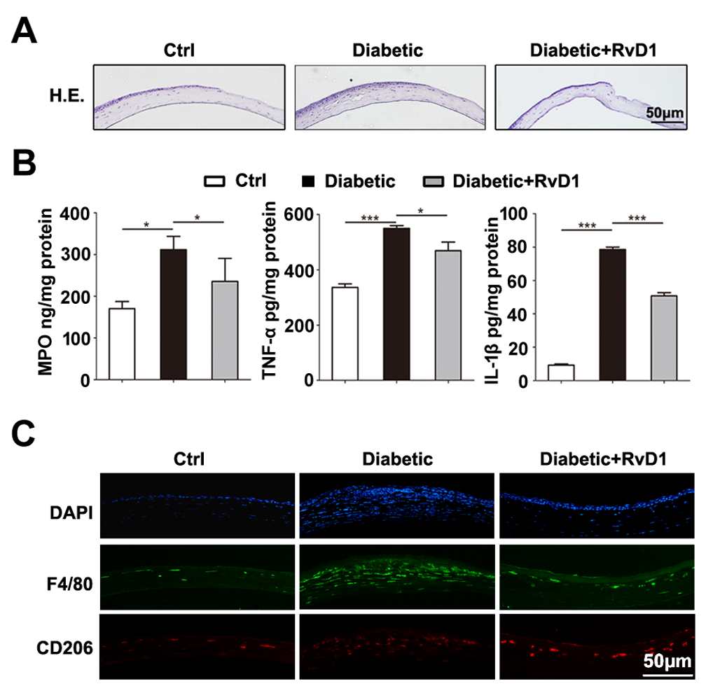
 Figure 3 of
Zhang, Mol Vis 2018; 24:274-285.
Figure 3 of
Zhang, Mol Vis 2018; 24:274-285.