Figure 2. Clinical photographs of the eyes of individuals with aniridia. A: Slit-lamp photo showing total aniridia (individual 5). B: Slit-lamp retroillumination photo showing total aniridia (individual 2). C: Slit-lamp retroillumination photo showing polar cataract (individual 5). D: Slit-lamp retroillumination photo showing anterior polar and cortical cataract (individual 7). E: Slit-lamp retroillumination photo showing lens subluxation (individual 1b). F: Slit-lamp photo showing opacification and vascularization of the cornea due to limbal stem cell failure (individual 1b).
G: Fundus photography showing small and tilted disc (individual 2). H: Fundus photography showing macular hypoplasia (individual 2).
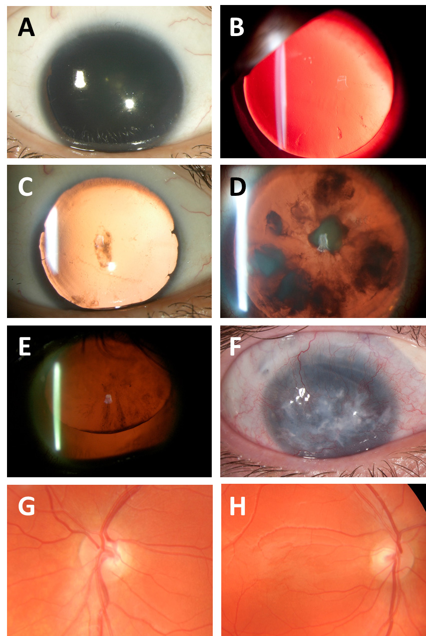
 Figure 2 of
Souzeau, Mol Vis 2018; 24:261-273.
Figure 2 of
Souzeau, Mol Vis 2018; 24:261-273.