Figure 2. Changes in morphology and location of IBA1-positive microglia and macrophages in ob/ob mice. A–C: There was no change in the overall numbers of IBA1-positive cells between the ob/ob retinas and the wild-type (wt) control
retinas (A), with an occasional IBA1-positive cell process found in the outer nuclear layer (ONL) in the wt control mice (B) and ob/ob (C) mice. D–G: However, several IBA1-positive cells were found with nuclei in the outer retina in the ob/ob mice only, with processes extending
into the outer segments (OS, D–F) and RPE (G). No IBA1-positive nuclei were observed in the outer retinas (ONL-RPE) of the wt control animals. Statistical analysis was
performed using a Student t test. NS indicates no statistical significance (p>0.05). GCL, ganglion cell layer; INL, inner nuclear layer. Scale bars =
50 µm. Error bars are displayed as standard error of the mean (SEM).
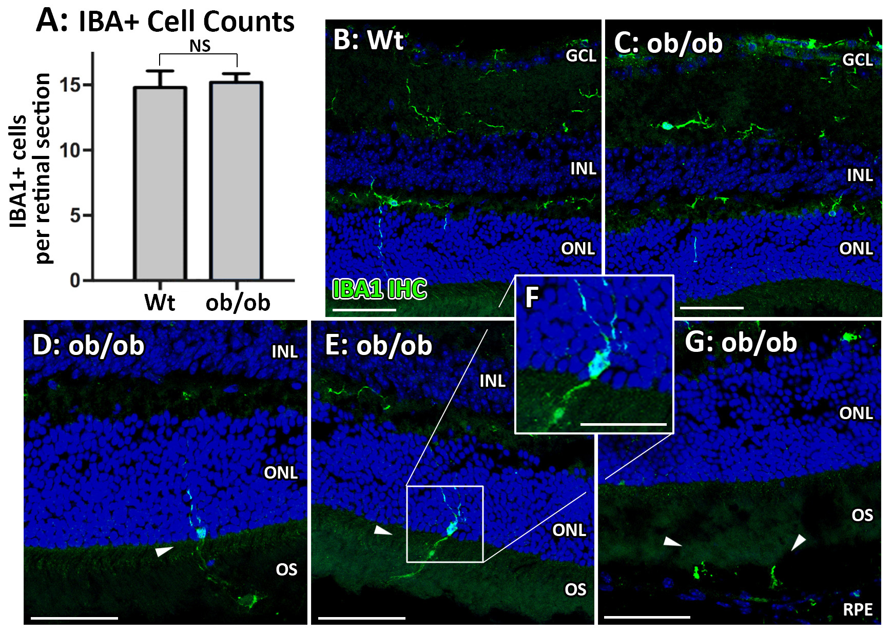
 Figure 2 of
Natoli, Mol Vis 2018; 24:201-217.
Figure 2 of
Natoli, Mol Vis 2018; 24:201-217.