 Appendix 1 of
Natoli, Mol Vis 2018; 24:201-217.
Appendix 1 of
Natoli, Mol Vis 2018; 24:201-217. Appendix 1 of
Natoli, Mol Vis 2018; 24:201-217.
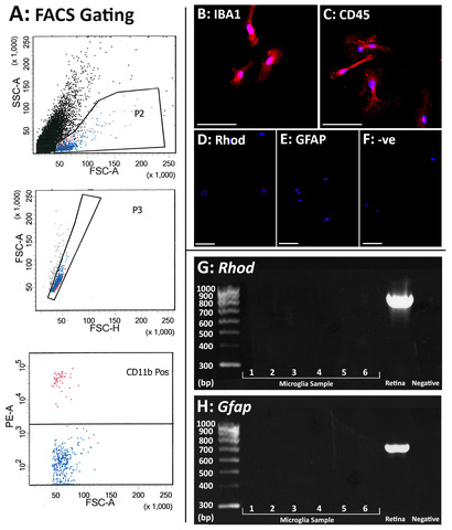
Appendix 1. Validation of primary isolated microglia as pure cultures.
A: Representative plots of the gating strategy used in fluorescence-activated cell sorting (FACS) to isolate CD11b+ cells (red population) from whole retinas. B-F: Microglial cells were immunolabelled with monocyte markers IBA1 (B, red) and CD45 (C, red), with nuclei counterstained with DAPI. Immunolabelling for other common retinal cell markers including Rhodopsin (Rhod, D), GFAP (E) and a negative control (-ve, F) showed no positive labeling for these markers. G-H: Primary microglial cells were also screened for other common retinal cell markers including Rhod (G) and Gfap (H) using PCR. There was no expression in any of the isolated microglial cell samples for Rhod or Gfap, however positive PCR banding was detected in a whole retinal sample indicating successful PCR amplification. Scale bars indicate 50 µm. To access the data, click or select the words “Appendix 1”