Figure 3. Corneal images (A), clinical scores (B), and the mRNA levels of IFN-α, IFN-β, and IFN-γ (C) in the murine fungal keratitis model on day 1, day 3, day 5, and day 7 post-infection. C57BL/6 mice were infected with Aspergillus fumigatus following routine protocols. The corneal images (A) and clinical scores (B) indicate the characteristics of corneal lesions at day 1, day 3, and day 5 post-infection. The mRNA levels of interferon
(IFN)-α, IFN-β, and IFN-γ (normalized to β-actin) increased in a time-dependent manner, peaking on day 5 post-infection, and
then were reduced on day 7 (C to E). Magnification: 16X. The data are the mean ± standard error of the mean (SEM) and represent three individual experiments
with six animals/group/time. *, p<0.05; **, p<0.01; ***, p<0.001.
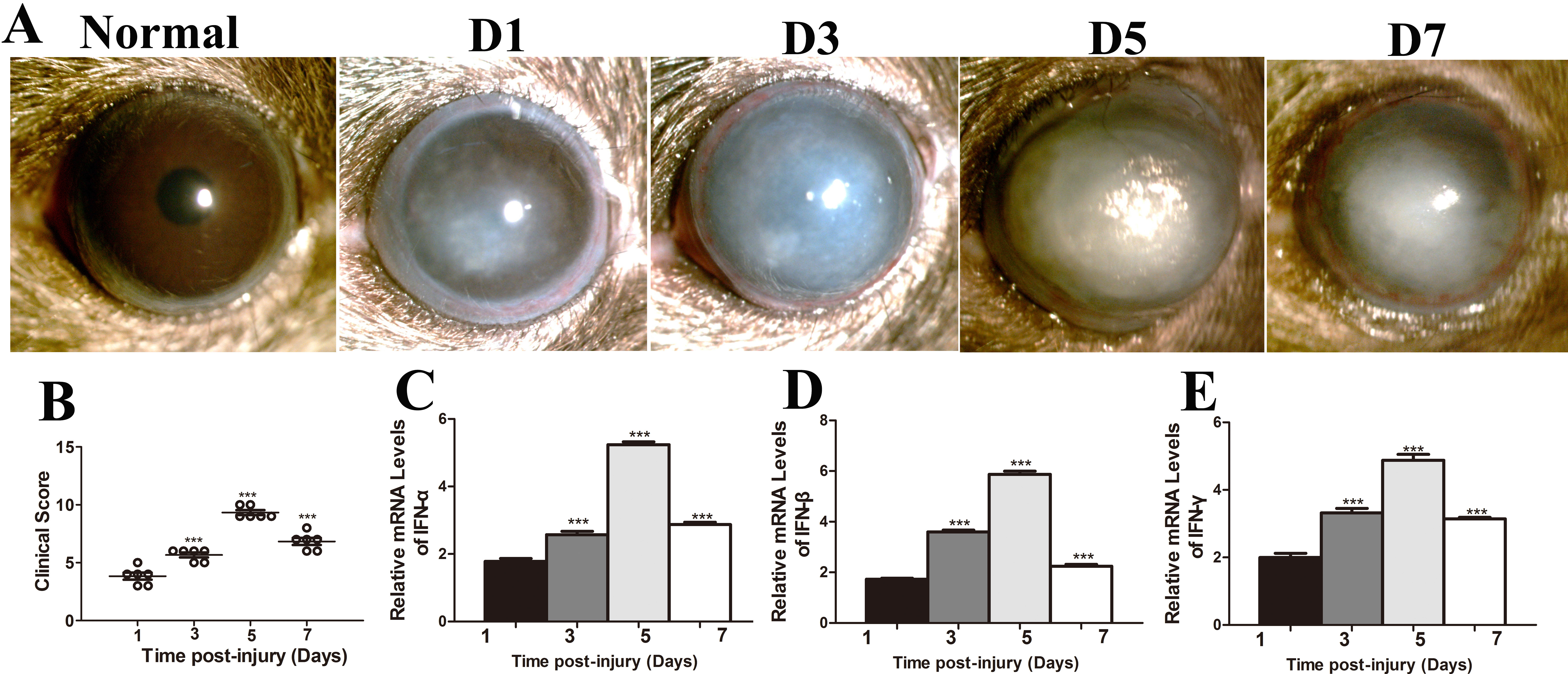
 Figure 3 of
Zhong, Mol Vis 2018; 24:187-200.
Figure 3 of
Zhong, Mol Vis 2018; 24:187-200.