Figure 3. M-cone ERG responses and human M- and L-opsin expression in AAV5-PR2.1-OPN1MW- or AAV5-PR2.1-OPN1LW-treated Opn1mw−/− eyes. A: M-cone electroretinogram (ERG) responses from untreated (UTx), OPN1MW-treated, and OPN1LW-treated Opn1mw-/ eyes and wild-type controls. Each bar represents the mean ± standard error of the mean (SEM) of M-cone b-wave amplitudes
recorded at 1.4 log cd.s/m2 (n = 5 for each group, *p<0.005). B: Western blot analysis of OPN1MW and OPN1LW expression in untreated Opn1mw−/− eyes (UTx), OPN1MW-, or OPN1LW- treated Opn1mw−/− eyes and wild-type (WT) untreated control eyes. C: Immunohistochemistry of AAV–mediated OPN1MW and OPN1LW expression in the treated Opn1mw−/− retinas. In the untreated eyes, no M-opsin expression was detected. S-opsin (green) was expressed normally in the ventral
retina (bottom row, left panel). The peanut agglutinin (PNA) staining (green) was normal in the dorsal and ventral retinas
(top and bottom rows, second column from the left). In the treated eyes, AAV-delivered OPN1MW and OPN1LW were detected in
the dorsal and ventral retinas. The vectored opsins were colocalized with PNA in the dorsal retinas (top row, two right panels)
and with endogenous S-opsin in the ventral retinas (bottom row, two right panels). Scale bar: 20 µm.
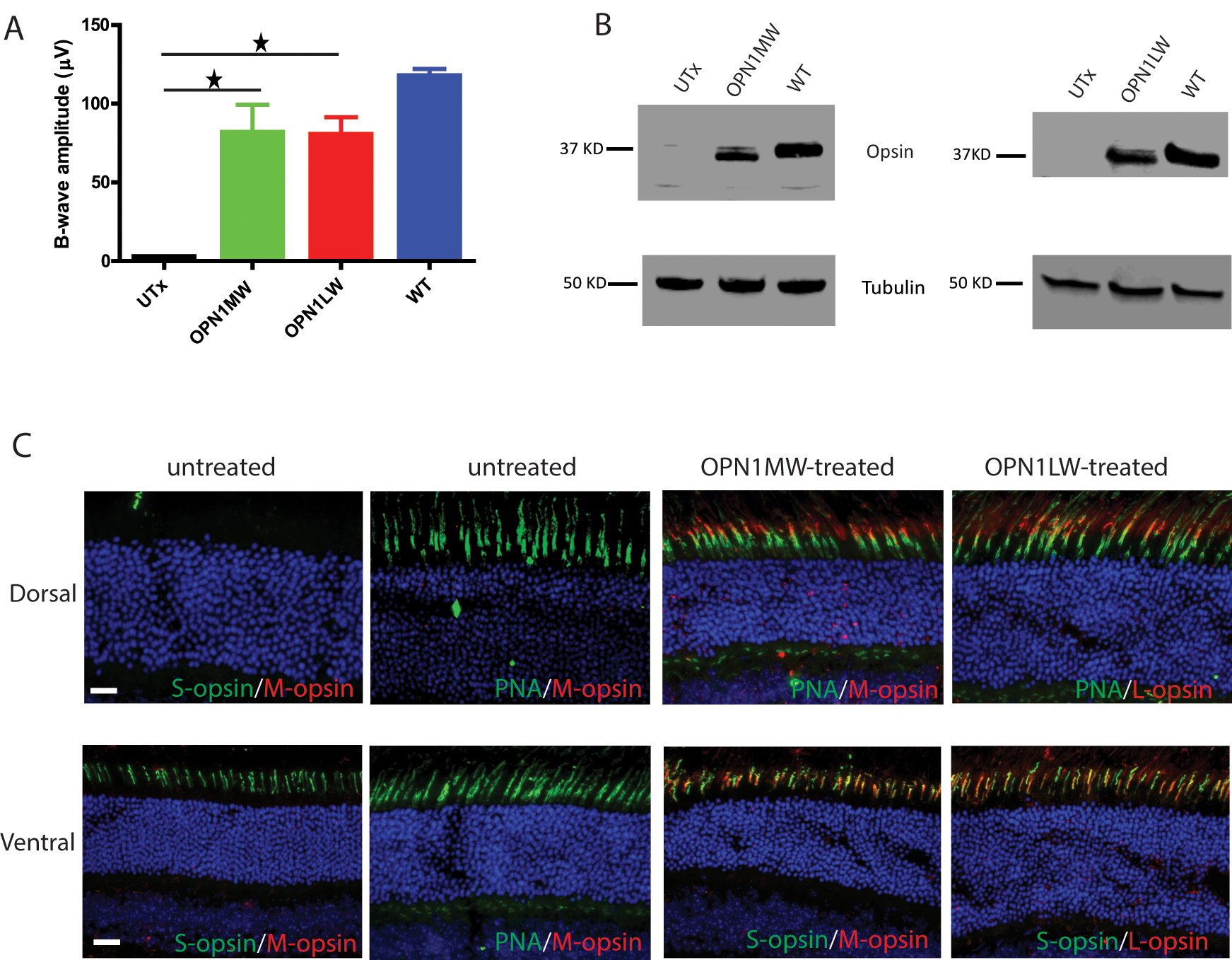
 Figure 3 of
Deng, Mol Vis 2018; 24:17-28.
Figure 3 of
Deng, Mol Vis 2018; 24:17-28.