Figure 2. Cone outer segments are restored in the treated Opn1mw−/− dorsal retinas. A: Images from untreated, OPN1MW- or OPN1LW-treated Opn1mw−/− dorsal retinas labeled with PDE6α’ (top row) or cTα (bottom row) showing that cone outer segments were regenerated in the
treated retinas. Adeno-associated virus (AAV)–delivered human opsins (red) were observed only in treated eyes, and the human
opsins were colocalized with PDE6α’ and cTα in the cone outer segments. Scale bar: 20 µm. B: An image from the dorsal-ventral transition zone in an OPN1LW-treated Opn1mw−/− eye showing equal lengths of cone outer segments with PDE6α’ staining. S-opsin was detected only in the ventral hemisphere.
C: Normalized average cone outer and inner segment lengths visualized with PDE6α’ staining (left panel), and normalized average
cone outer and inner segment sheath lengths visualized with peanut agglutinin (PNA) staining (right panel) from the untreated
Opn1mw−/− dorsal retinas (uTx dorsal), OPN1LW-treated Opn1mw−/− dorsal retinas (Tx dorsal), untreated Opn1mw−/− ventral retinas (uTx ventral), and wild-type controls (WT dorsal, WT ventral). Each bar represents the average of 90 cones
from three images each from three individual eyes of different animals. Error bars represent the standard error of the mean
(SEM; *p<0.001). COS: cone outer segments; CIS: cone inner segments.
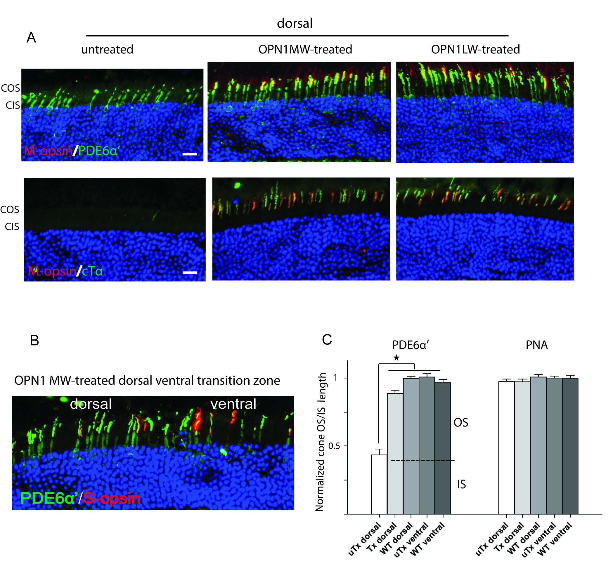
 Figure 2 of
Deng, Mol Vis 2018; 24:17-28.
Figure 2 of
Deng, Mol Vis 2018; 24:17-28.