Figure 1. Cone outer segments are shortened in dorsal retinas of Opn1mw−/− mice. A: PDE6α’ staining (top row) in Opn1mw−/− dorsal cones is observed only in the inner segments, while its staining in ventral cones is concentrated in outer segments
shown as colocalization with endogenous S-opsin. PDE6α’ staining is primarily observed in cone outer segments in the wild-type
control. cTα staining (bottom row) is absent in the dorsal cones of the Opn1mw−/− retina, while staining is present in cone outer segments in Opn1mw−/− ventral cones as in the wild-type control. As expected, S-opsin staining (red) was primarily observed in ventral retinas
in Opn1mw−/− and wild-type mice. B: An image from the Opn1mw−/− dorsal-ventral transition zone showing shortened cone outer segments in the dorsal hemisphere with PDE6α’ staining (left
panel). The right panel is the same image superimposed with peanut agglutinin (PNA) staining showing equal lengths of cone
outer and inner segment sheaths between the dorsal and ventral hemispheres. COS: cone outer segments; CIS: cone inner segments.
Scale bar: 20 µm.
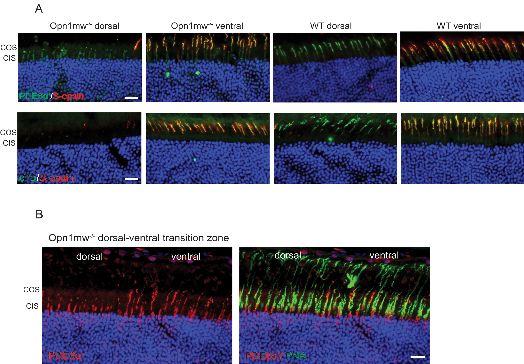
 Figure 1 of
Deng, Mol Vis 2018; 24:17-28.
Figure 1 of
Deng, Mol Vis 2018; 24:17-28.