Figure 4. Expression of cyclin D1 in ACCs in the lacrimal gland. A: Healthy lacrimal glands. B: Tubular pattern. C: Cribriform pattern. D: Solid pattern. Cyclin D1 expression was positive in all cases. Red curves: inner ductal epithelial cells; black arrows:
positive staining of cyclin D1. Scale bars = 50 μm.
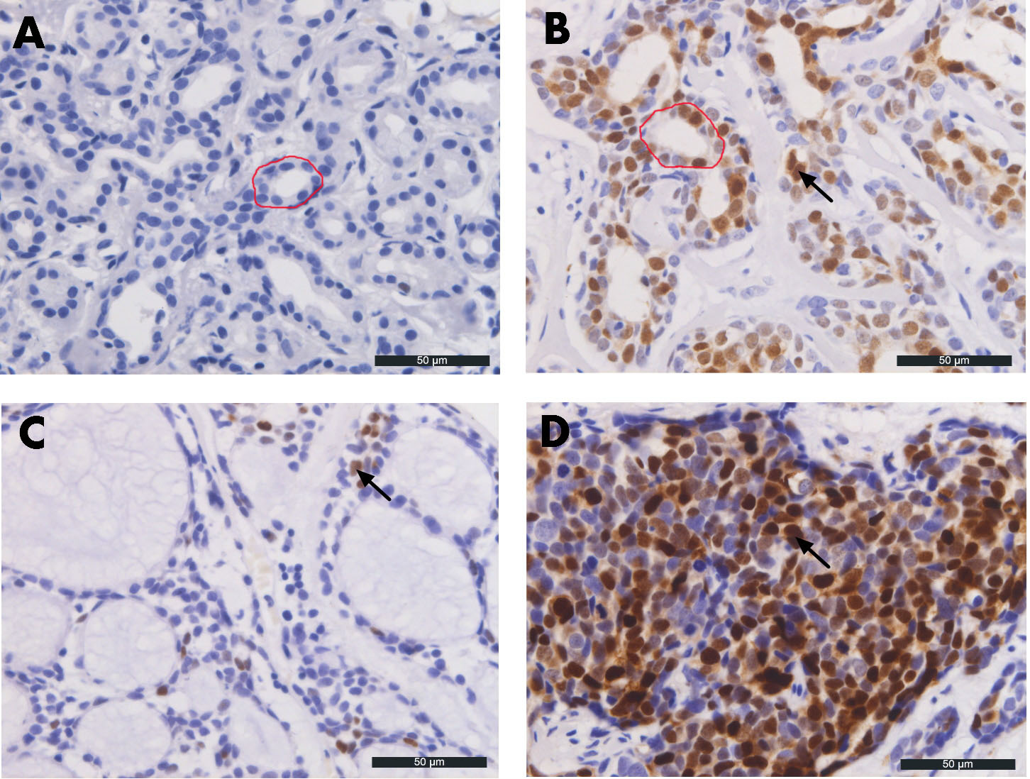
 Figure 4 of
Wang, Mol Vis 2018; 24:143-152.
Figure 4 of
Wang, Mol Vis 2018; 24:143-152.