Figure 3. Retinal structure in patients with the p.Ala1773Val mutation in ABCA4. A–D: Cross-sectional optical coherence tomography (OCT) images along the horizontal meridian through the fovea in a healthy subject
compared with three patients with Stargardt disease (STGD1; P2, P6, and P7) with different patterns of central retinal degeneration.
A:Retina from a 28-year-old healthy subject with normal central lamination. Magnified OCT images from the extrafoveolar regions
delineated by white rectangles on the images illustrate the anatomy of the inner and outer retina. B:Retina from a 26-year-old patient with central retinal degeneration where the ONL, ELM, and EZ line are detectable along the
central OCT scan. C: Retina from a 32-year-old patient with central retinal degeneration and a detectable ONL and ELM along the central OCT scan.
D: Retina from a 30-year-old patient with central retinal degeneration and only the ONL detectable along the central OCT scan.
The white arrow points to the outer nuclear layer. The dashed line with the white arrow points to the ELM. The white arrowhead
points to the EZ line. ONL; outer nuclear layer. ELM; external limiting membrane. EZ; ellipsoid zone. E–G: Short-wave autofluorescence imaging results of a retina from a healthy subject compared with two representative patterns
of abnormal autofluorescence imaging in two patients with STGD1 (P2 and P6) with ABCA4 retinopathy. E: SW-AF of a retina from a healthy 28-year-old male shows a central lower autofluorescence signal that increases eccentrically.
The blood vessels and optic nerve appear dark. F:Retina from a 26-year-old patient with STGD1 shows a central hypoautofluorescence surrounded by a heterogeneous signal extending
anterior to the vascular arcades. G:Retina from a 30-year-old patient with STGD1 shows a central hypoautofluorescence signal surrounded by patches of decreased
fluorescence.
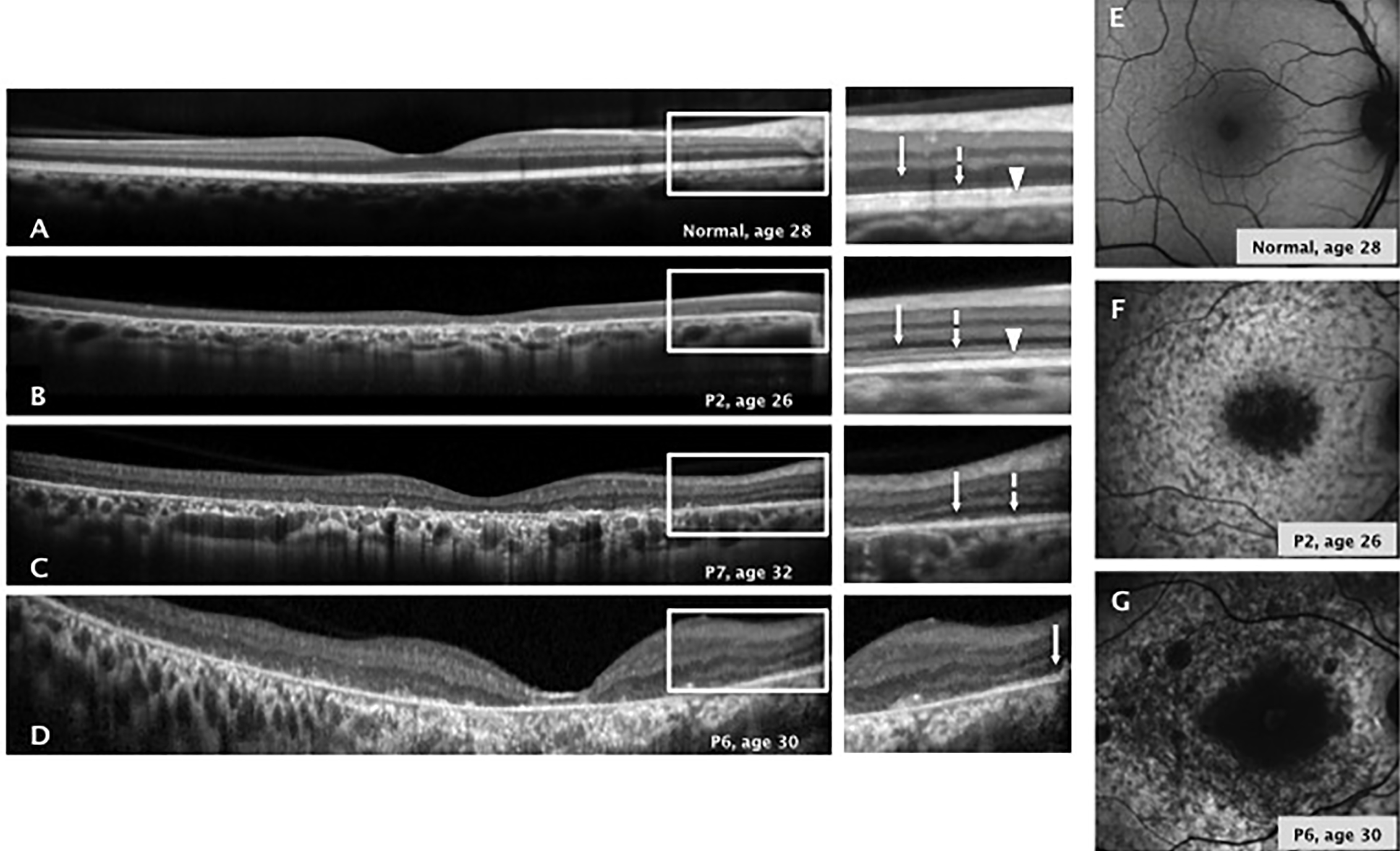
 Figure 3 of
López-Rubio, Mol Vis 2018; 24:105-114.
Figure 3 of
López-Rubio, Mol Vis 2018; 24:105-114.