Figure 4 of
Sun, Mol Vis 2017; 23:977-986.
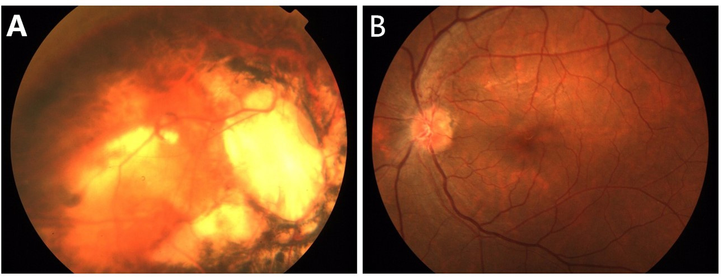
Figure 4.
Fundus photographs of simplex case 2.
A
: Right eye.
B
: Left eye. The fundus photographs of simplex case 2 show optic disc coloboma for the right eye and a relatively normal phenotype for the left eye.
 Figure 4 of
Sun, Mol Vis 2017; 23:977-986.
Figure 4 of
Sun, Mol Vis 2017; 23:977-986.