Figure 3. B-ultrasonography images of patients from family B. A and B: Images of the right and left eyes, respectively of patient II:2. C and D: Images of the right and left eyes, respectively, of patient III:1. The B-ultrasonography images show posterior segment coloboma
for both eyes of patient II:2 and the right eye of patient III:1. Focal and irregular introcessions in the eyeball wall are
marked with red arrows.
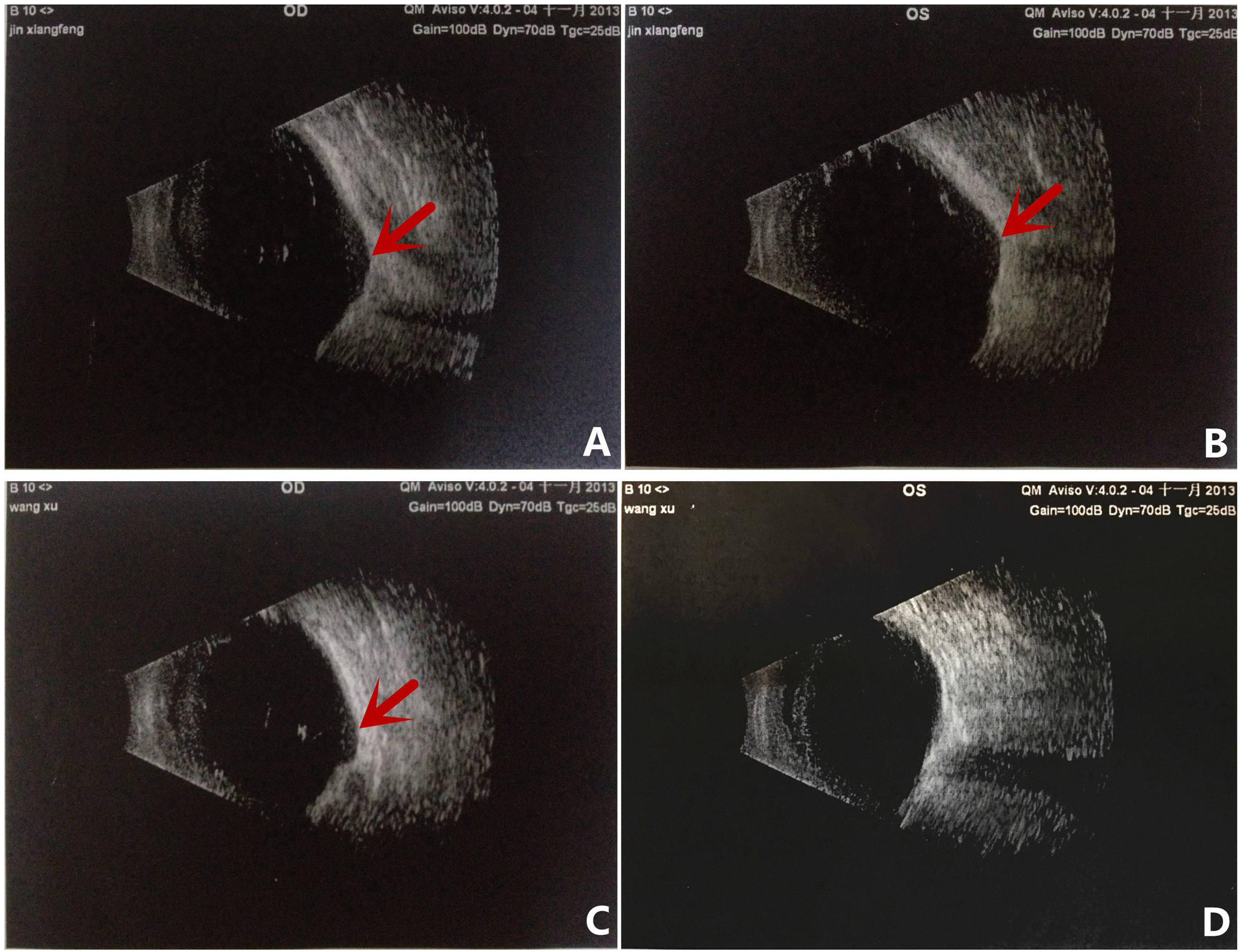
 Figure 3 of
Sun, Mol Vis 2017; 23:977-986.
Figure 3 of
Sun, Mol Vis 2017; 23:977-986.