Figure 5. Changes in NG2-positive/OPCs in the anterior ON 30 day post-rNAION. A, B: Optic nerve (ON) cross sections from A) naïve and B) rodent nonarteritic anterior ischemic optic neuropathy (rNAION)-induced. There is a considerable increase in the number
of oligodendrocyte progenitor cells (OPCs) present in the ON following ON ischemia. C: Quantification of NG2-positive/OPC increase 30 days post-rNAION. There is a 7.1-fold increase in the number of NG2-positive
cells present in the rNAION-induced nerve (28.7±2.70 v. 4.0±1.9 (SEM) cells/field of view (FOV; n = 6 animals); ANOVA; f-ratio
= 55.09054, p=0.000023.
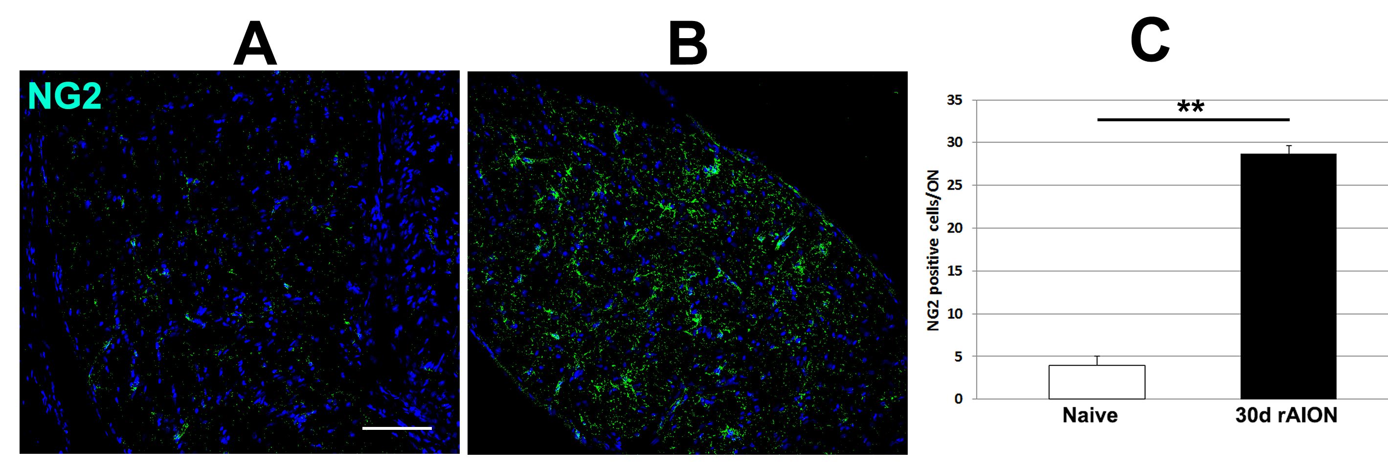
 Figure 5 of
Mehrabian, Mol Vis 2017; 23:963-976.
Figure 5 of
Mehrabian, Mol Vis 2017; 23:963-976.