Figure 3. SP-induced suppression of local inflammation contributes to the inhibition of cellular apoptosis and α-SMA (+) cell infiltration
at wound sites. A,B: Immunohistochemical staining for tumor necrosis factor-alpha (TNF-α) was performed, and the intensity of the stained area
was measured using the ImageJ program. C,D: Analysis of the apoptotic cells in the retina was performed. To elucidate the apoptotic cells, cleaved caspase 3 (+) cells
were counted in the inner nuclear layer (INL). Red arrow: cleaved caspase 3 (+) cells. E,F: Alpha smooth muscle actin (α-SMA) (+) fibroblastic cells migrated toward the INL were observed and quantitatively assessed.
G: Level of TNF-α, cleaved caspase-3, and α-SMA were determined by western blotting. The mean measurement of the six images
in each tissue sample was calculated in each animal. Data are presented as mean ± standard deviation (SD). Scale bar: 50 μm.
*p<0.05; **p<0.01; n = 8 mice per group.
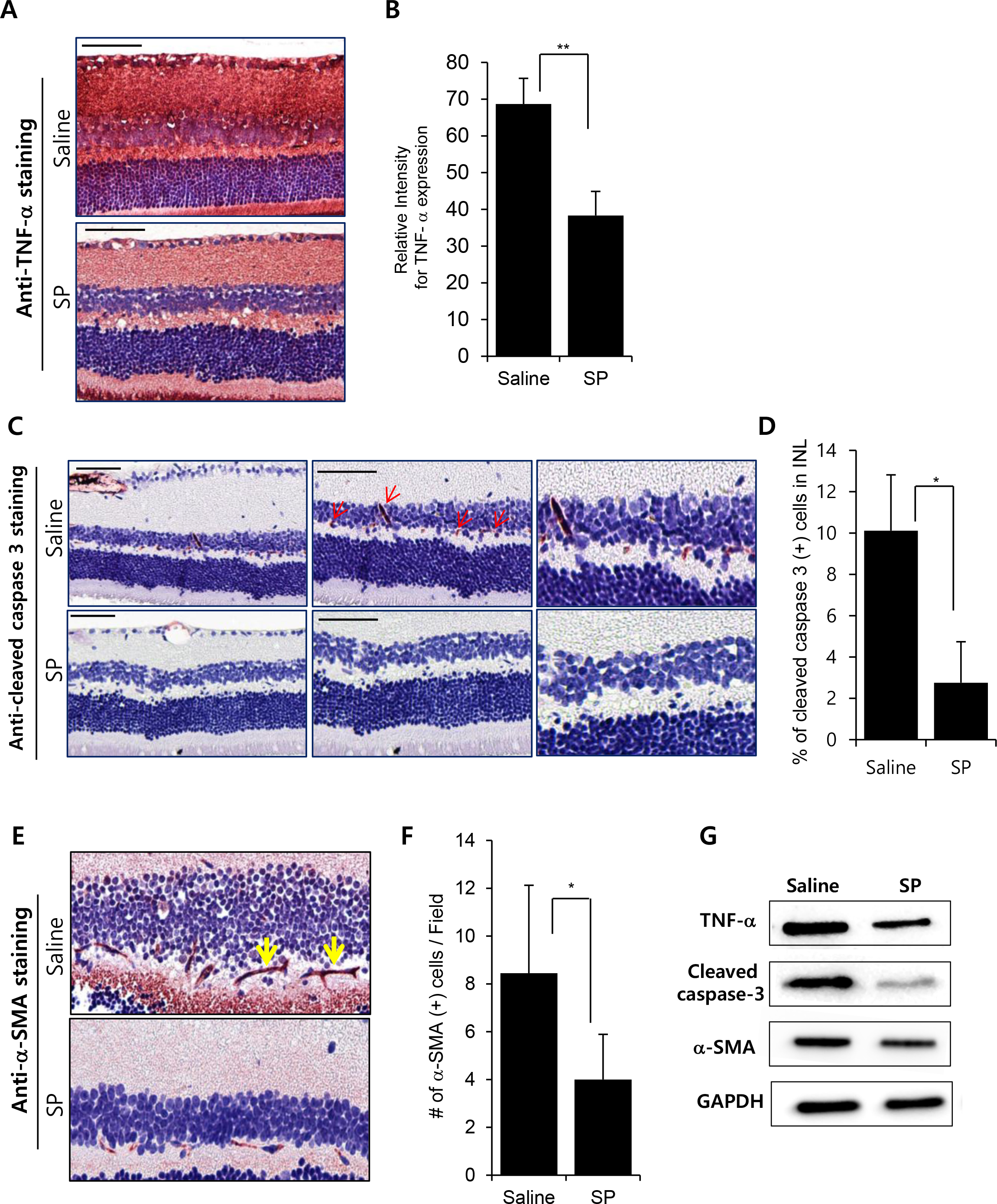
 Figure 3 of
Yoo, Mol Vis 2017; 23:933-943.
Figure 3 of
Yoo, Mol Vis 2017; 23:933-943.