Figure 2. Neutrophils infiltration into the anterior and posterior chambers of the eye. Sham-injected (A, D), interleukin (IL)-8 (B, E) and tumor necrosis factor alpha (TNF-α) (C, F), 24 h post-injection. No cellular infiltration was observed in the sham-injected eyes (A, E) while abundant CD11b+ cells were seen in the eyes injected with IL-8 (B, E), and TNF-α (C, F). C= cornea; AC = anterior chamber; V = vitreous; GCL = ganglion cell layer. The results show representative images observed
in five eyes (n=5/group). Magnification = 63X. Scale bar = 20 μm.
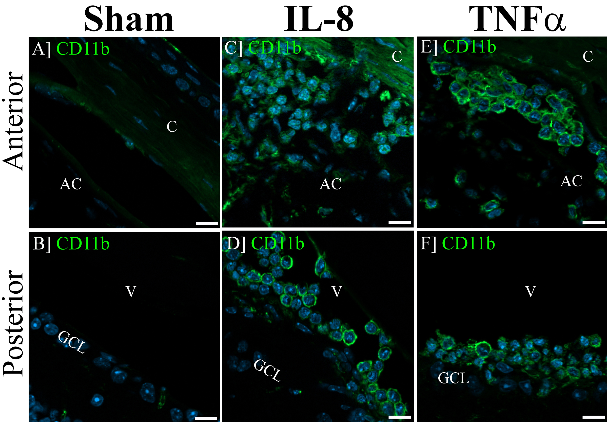
 Figure 2 of
Barliya, Mol Vis 2017; 23:922-932.
Figure 2 of
Barliya, Mol Vis 2017; 23:922-932.